. 2024 Mar 27;10(1):155.
doi: 10.1038/s41420-024-01925-4.

METTL3-mediated m6A modification increases Hspa1a stability to inhibit osteoblast aging

Affiliations

METTL3-mediated m6A modification increases Hspa1a stability to inhibit osteoblast aging

Yaobin Wang et al. Cell Death Discov. .

Abstract

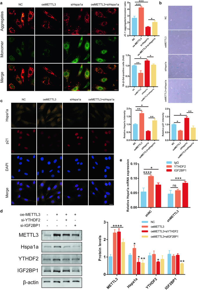
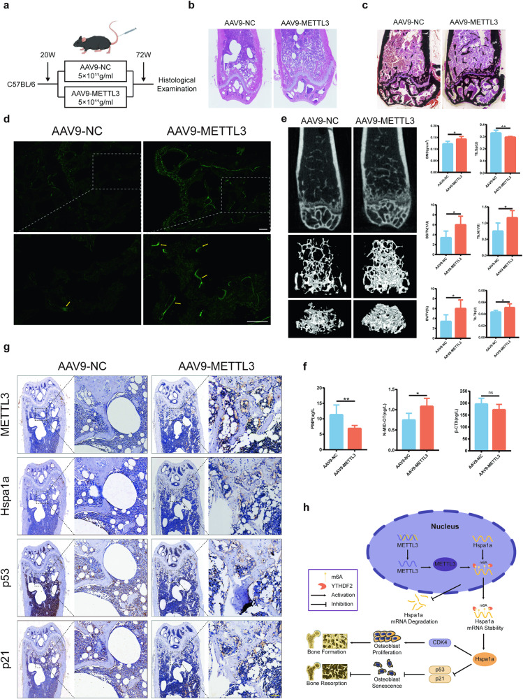
Senile osteoporosis is mainly caused by osteoblasts attenuation, which results in reduced bone mass and disrupted bone remodeling. Numerous studies have focused on the regulatory role of m6A modification in osteoporosis; however, most of the studies have investigated the differentiation of bone marrow mesenchymal stem cells (BMSCs), while the direct regulatory mechanism of m6A on osteoblasts remains unknown. This study revealed that the progression of senile osteoporosis is closely related to the downregulation of m6A modification and methyltransferase-like 3 (METTL3). Overexpression of METTL3 inhibits osteoblast aging. Methylated RNA immunoprecipitation sequencing (MeRIP-seq) revealed that METTL3 upregulates the stability of Hspa1a mRNA, thereby inhibiting osteoblast aging. Moreover, the results demonstrated that METTL3 enhances the stability of Hspa1a mRNA via m6A modification to regulate osteoblast aging. Notably, YTH N6-methyladenosine RNA binding protein 2 (YTHDF2) participates in stabilizing Hspa1a mRNA in the METTL3-mediated m6A modification process, rather than the well-known degradation function. Mechanistically, METTL3 increases the stability of Hspa1a mRNA in a YTHDF2-dependent manner to inhibit osteoblast aging. Our results confirmed the significant role of METTL3 in osteoblast aging and suggested that METTL3 could be a potential therapeutic target for senile osteoporosis.


Conflict of interest statement

The authors declare no competing interests.

Figures

Fig. 1
Fig. 1. Diminished m6A modification in osteoporosis in the elderly.
a Colorimetric assessment of m6A levels in bone samples from elderly osteoporosis patients (n = 10). b qRT-PCR analysis of METTL3, METTL14, FTO, and YTHDC1 expression (n = 10). c Colorimetric evaluation of m6A levels in mouse osteoporotic bone tissue (n = 6). d Western blotting to gauge METTL3 protein levels (n = 6). e Colorimetric analysis of m6A levels in H2O2-induced aging of MC3T3-E1 cells (n = 3). f Identification of senescent cells via β-galactosidase staining (n = 3). g Transmission electron microscopy for mitochondrial morphology (n = 3). h METTL3 protein levels were assessed by Western blotting (n = 3). Data represented as mean ± standard deviation from three independent experiments. *p < 0.05, **p < 0.01, ***p < 0.001, ****p < 0.0001 by Student’s t-test.
Fig. 2
Fig. 2. Osteoblast senescence was strongly linked to METTL3-facilitated m6A alterations.
a Colorimetric assessment of m6A levels in MC3T3-E1 and primary osteoblasts post-METTL3 knockout (n = 3). b β-Galactosidase staining to identify senescent cells (n = 3). c Confocal microscopy to reveal METTL3 and p21 localization and expression, bar = 20 μm (n = 3). d JC-1 staining to measure mitochondrial membrane potential, bar=10 μm (n = 3). e Western blotting for METTL3, p53, CDK4, and p21 protein levels (n = 3). **p < 0.01, ***p < 0.001, ****p < 0.0001, ns, no significant, by Student’s t-test.
Fig. 3
Fig. 3. Osteoblast function affects bone microenvironment.
a Confocal microscopy scans for METTL3 and p21 expression, bar = 20 μm (n = 3). b β-Galactosidase staining for senescent cells (n = 3). c JC-1 staining to assess mitochondrial membrane potential, bar = 10 μm (n = 3). d β-Galactosidase staining for senescent cells (n = 3). e EdU assay for cellular proliferation, bar = 50 μm (n = 3). *p < 0.05, **p < 0.01, ***p < 0.001, ****p < 0.0001 by Student’s t-test.
Fig. 4
Fig. 4. MeRIP-seq reveals that METTL3 regulates the stability of Hspa1a.
a MeRIP-Seq analysis showed “GGAC” motif enrichment in osteoblasts. b qRT-PCR analysis of seven significantly upregulated potential targets (n = 3). c High m6A peak abundance and specificity near the Hspa1a stop codon. d Western blotting to assess Hspa1a protein levels following METTL3 silencing (n = 3). e Confocal microscopy scans for Hspa1a expression, bar = 20 μm (n = 3). f RIP-qPCR to investigate direct Hspa1a mRNA-METTL3 protein interactions (n = 3). g MeRIP-qPCR to explore Hspa1a m6A modifications (n = 3). h qRT-PCR to measure mRNA degradation after actinomycin D induction (n = 3). *p < 0.05, **p < 0.01, ***p < 0.001, ****p < 0.0001 by Student’s t-test.
Fig. 5
Fig. 5. METTL3-driven m6A modification directs YTHDF2-dependent Hspa1a mRNA decay.
a JC-1 staining to measure mitochondrial membrane potential, bar = 10 μm (n = 3). b β-Galactosidase staining to identify senescent cells (n = 3). c Confocal microscopy scans for Hspa1a and p21 expression, bar = 20 μm (n = 3). d Western blotting for METTL3, Hspa1a, YTHDF2, and IGF2BP1 proteins (n = 3). e RIP-qPCR to identify m6A-dependent proteins interacting with Hspa1a mRNA (n = 3). *p < 0.05, **p < 0.01, ***p < 0.001, ****p < 0.0001, ns, no significant, by Student’s t-test.
Fig. 6
Fig. 6. METTL3 targeting in osteoblasts inhibits osteoporosis in aged mice.
a Diagrammatic representation of mouse groups and interventions (n = 6). b, c, f Histological evaluations (HE staining, Von Kossa staining, and calcein staining, bar = 200 μm) of the distal femur (n = 3). d Quantitative micro-CT analysis of the distal femur (n = 3). e ELISA for serum bone metabolic markers (n = 3). g Immunohistochemistry for METTL3, Hspa1a, p53, and p21 levels in bone tissue (n = 3). h Schematic diagram showing METTL3-mediated m6A modification regulates Hspa1a mRNA decay in a YTHDF2-dependent manner, and METTL3 controls the progression of osteoporosis in the elderly by inhibiting bone absorption through osteoblast senescence. bar = 50 μm. *p < 0.05, **p < 0.01, ns, no significant, by Student’s t-test.

Similar articles

Cited by

References

    1. Khosla S, Farr JN, Tchkonia T, Kirkland JL. The role of cellular senescence in ageing and endocrine disease. Nat Rev Endocrinol. 2020;16:263–75. doi: 10.1038/s41574-020-0335-y. - DOI - PMC - PubMed
    1. Khosla S, Farr JN, Monroe DG. Cellular senescence and the skeleton: pathophysiology and therapeutic implications. J Clin Investig. 2022;132:e154888. doi: 10.1172/JCI154888. - DOI - PMC - PubMed
    1. Cui J, Shibata Y, Zhu T, Zhou J, Zhang J. Osteocytes in bone aging: Advances, challenges, and future perspectives. Ageing Res Rev. 2022;77:101608. doi: 10.1016/j.arr.2022.101608. - DOI - PubMed
    1. Farr JN, Xu M, Weivoda MM, Monroe DG, Fraser DG, Onken JL, et al. Targeting cellular senescence prevents age-related bone loss in mice. Nat Med. 2017;23:1072–9. doi: 10.1038/nm.4385. - DOI - PMC - PubMed
    1. Wang Y, Deng P, Liu Y, Wu Y, Chen Y, Guo Y, et al. Alpha-ketoglutarate ameliorates age-related osteoporosis via regulating histone methylations. Nat Commun. 2020;11:5596. doi: 10.1038/s41467-020-19360-1. - DOI - PMC - PubMed