An official website of the United States government
The .gov means it’s official.
Federal government websites often end in .gov or .mil. Before
sharing sensitive information, make sure you’re on a federal
government site.
The site is secure.
The https:// ensures that you are connecting to the
official website and that any information you provide is encrypted
and transmitted securely.
Radko-Juettner S, Yue H, Myers JA, Carter RD, Robertson AN, Mittal P, Zhu Z, Hansen BS, Donovan KA, Hunkeler M, Rosikiewicz W, Wu Z, McReynolds MG, Roy Burman SS, Schmoker AM, Mageed N, Brown SA, Mobley RJ, Partridge JF, Stewart EA, Pruett-Miller SM, Nabet B, Peng J, Gray NS, Fischer ES, Roberts CWM.Radko-Juettner S, et al.Nature. 2024 May;629(8012):E12. doi: 10.1038/s41586-024-07402-3.Nature. 2024.PMID: 38684813No abstract available.
Abstract
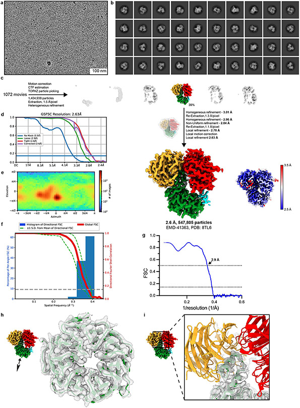
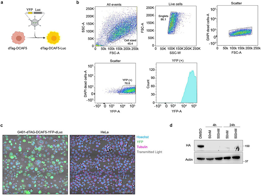
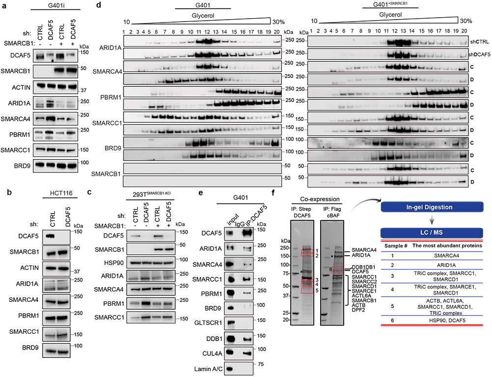
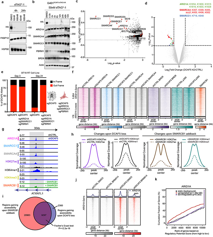
Whereas oncogenes can potentially be inhibited with small molecules, the loss of tumour suppressors is more common and is problematic because the tumour-suppressor proteins are no longer present to be targeted. Notable examples include SMARCB1-mutant cancers, which are highly lethal malignancies driven by the inactivation of a subunit of SWI/SNF (also known as BAF) chromatin-remodelling complexes. Here, to generate mechanistic insights into the consequences of SMARCB1 mutation and to identify vulnerabilities, we contributed 14 SMARCB1-mutant cell lines to a near genome-wide CRISPR screen as part of the Cancer Dependency Map Project1-3. We report that the little-studied gene DDB1-CUL4-associated factor 5 (DCAF5) is required for the survival of SMARCB1-mutant cancers. We show that DCAF5 has a quality-control function for SWI/SNF complexes and promotes the degradation of incompletely assembled SWI/SNF complexes in the absence of SMARCB1. After depletion of DCAF5, SMARCB1-deficient SWI/SNF complexes reaccumulate, bind to target loci and restore SWI/SNF-mediated gene expression to levels that are sufficient to reverse the cancer state, including in vivo. Consequently, cancer results not from the loss of SMARCB1 function per se, but rather from DCAF5-mediated degradation of SWI/SNF complexes. These data indicate that therapeutic targeting of ubiquitin-mediated quality-control factors may effectively reverse the malignant state of some cancers driven by disruption of tumour suppressor complexes.
Extended Data Figure 1.. DCAF5 dependence is specific to SMARCB1 -mutant cancers independent of mRNA…
Extended Data Figure 1.. DCAF5 dependence is specific to SMARCB1-mutant cancers independent of mRNA expression and tissue type.
a, Two-class comparison of n=14 biologically independent Rhabdoid Tumor cell lines compared to other SWI/SNF mutant cancer cell lines (n=190) or other non-SWI/SNF mutant cancer cell lines (n=607) (****P = 1.22 x 10⁻¹⁶ and 7.16 x 10⁻²¹ respectively; two-tailed Student’s t test, release CERES 21Q1). The box plot indicates the median (center line), the third and first quartiles (box limits) and 1.5 × interquartile range (IQR) above and below the box (whiskers). b, Box plot showing DCAF5 RNA expression across n= 1332 biologically independent cancer cell lines from different tumor types in the Cancer Cell Line Encyclopedia (CCLE) database. RT cell lines are shaded in red. The box plot indicates the median (center line), the third and first quartiles (box limits) and 1.5 × interquartile range (IQR) above and below the box (whiskers). c, Bar plot demonstrating normalized expression (nTPM) levels of DCAF5 for n=55 tissue types, created by combining the HPA and GTEx transcriptomics datasets using the Human Protein Atlas internal normalization pipeline. Color-coding is based on tissue groups. d, Effects on proliferation upon DCAF5 shRNA knockdown in SMARCB1-mutant cell lines. Solid lines (shCTRL) and dotted lines (shDCAF5). Graphs show mean values from n=8 technical replicates per cell line condition from one independent biological experiment. e, Western blot analysis of TTC549 RT cell line at Day 0 and Day 8 of IncuCyte proliferation assay. Band intensities were quantified by the Licor Image Studio Lite software and then the normalized DCAF5 level was calculated relative to Actin and normalized to shControl signal ± s.e.m (n=3 independent biological replicates). ***P= 0.001; Two-Way ANOVA. f, DCAF5 immunoprecipitation in G401 and BT16 RT cell line demonstrates interaction of DCAF5 with E3 Ub Ligase machinery. Input is 1% of the protein used for the IP (n=2 independent biological replicates).
Extended Data Figure 2.. Cryo-EM processing workflow…
Extended Data Figure 2.. Cryo-EM processing workflow for the DDB1∆B-DDA1-DCAF5 structure.
a , Raw micrograph…
Extended Data Figure 2.. Cryo-EM processing workflow for the DDB1∆B-DDA1-DCAF5 structure.
a, Raw micrograph (low pass-filtered to 10Å, scale bar indicated). b, Representative 2D classes. c, Overview of processing workflow from raw micrograph. All processing steps were conducted in cryoSPARC. Particles belonging to colored volumes were taken for the final map (EMD-41363). The final map is contoured at 0.134, and local resolution mapped onto the final reconstruction is shown. d, FSC plot for the deposited map (EMDB-41363). e, Viewing distribution plot. f, Directional resolution histogram and directional FSC plot. g, Model-to-map FSC for the deposited structure (PDB: 8TL6), value given for FSC (model)=0.5. h, Density example for the DCAF5 WD40 domain. i, Density for the DCAF5-motif in the DDB1ΔB binding site.
Extended Data Figure 3.. Details of DDB1∆B…
Extended Data Figure 3.. Details of DDB1∆B -DDA1-DCAF5 structure, evolutionary analysis and AlphaFold prediction.
a …
Extended Data Figure 3.. Details of DDB1∆B -DDA1-DCAF5 structure, evolutionary analysis and AlphaFold prediction.
a, Detailed view of DCAF5 and DDB1ΔB interaction shown in cartoon representation. The N-terminal α-helix of DCAF5 tightly inserts into the pocket of DDB1. b, Charge complementarity between DCAF5 and DDB1 at the interface. c, The N terminus of DDA1 inserts into DDB1, while the C terminus of DDA1 binds DCAF5 tightly with a hydrophobic interaction. DCAF5 surface is shown with hydrophobic and hydrophilic color coding. d, Plot of the ConSurf conservation score versus the amino acid residue of full-length DCAF5 with domain annotations. e, ConSurf conservation scores are mapped onto DCAF5 with orange-white-purple color scale in increasing conservation order. Top view and bottom view of the WD40 domain are shown. f, AlphaFold predictions for the DCAF5 aa 1-601 and SMARCC1 interaction. In the domain bar, DCAF5 is represented in green, with the WD40 domain specifically highlighted. SMARCC1 is depicted in magenta. g, The AlphaFold predicted binding mode of DCAF5 and SMARCC1 is shown. DCAF5 is represented in green, SMARCC1 is depicted in magenta, and DDB1-DDA1 is represented in grey.
Extended Data Figure 4.. DCAF5 loss upregulates…
Extended Data Figure 4.. DCAF5 loss upregulates protein levels of SWI/SNF members and alters SWI/SNF…
Extended Data Figure 4.. DCAF5 loss upregulates protein levels of SWI/SNF members and alters SWI/SNF complex integrity.
a, Western blot analysis of SWI/SNF subunits in TTC549 SMARCB1-inducible RT cells treated with shCTRL or shDCAF5 after 72 hours selection in the presence or absence of SMARCB1. b, RNA-Seq analysis in G401 RT cells treated with shCTRL or shDCAF5 after 72 hours selection evaluating log2 fold change of mRNA for SWI/SNF in shDCAF5 versus shCTRL. ns = not significant; ** log2FC = −0.68, FDR = 0.02. Significance was determined by two-sided Empirical Bayes test for differential expression with FDR adjusted p-values. c, Left: Cycloheximide Chase (0-24 hours with 50 ug/mL cycloheximide) in G401 shCTRL or shDCAF5 evaluating SWI/SNF subunit levels and control protein c-myc. Right: Graphical representation of the cycloheximide experimental data for the mean relative protein amount ± s.e.m of ARID1A (****P=<0.0001), SMARCA4 (**P=<0.0022), SMARCC1 (*P=<0.0180), PBRM1(****P=<0.0001) and c-myc (P= ns:not significant); Two-Way ANOVA. d, Glycerol gradient (10-30% glycerol) analysis of SMARCB1-deficient BT16 RT cells treated with either shCTRL or shDCAF5 after 72 hours selection (top panel). SMARCB1 has been re-expressed in the cells in the bottom panel. e, SMARCA4 co-immunoprecipitation in G401 shCTRL and shDCAF5 conditions demonstrates that the SWI/SNF complex is maintained in the absence of DCAF5. Lamin A/C is a negative control. Input is 1% of the protein used for the IP. f, SMARCA4 co-immunoprecipitation in G401-dTAG-DCAF5 cells treated with DMSO and V-1 demonstrates retained SWI/SNF complex interactions in the absence of DCAF5. Lamin A/C is a negative control. Input is 1% of the protein used for the IP. g, SMARCA4 co-immunoprecipitation in G401 RT cells demonstrates interaction with DCAF5 and SWI/SNF subunits. Lamin A/C is a negative control. Input is 1% of the protein used for the IP. h, Two-class comparison of n=14 biologically independent Rhabdoid Tumor cell lines compared to n=789 biologically independent other cancer cell lines in DepMap analyzing L3MBTL3 and LSD1 dependency (P = 0.907 and 0.701 respectively and is non-significant (ns); two-tailed Student’s t test, release CERES 21Q1). The box plots indicate the median (center line), the third and first quartiles (box limits) and 1.5 × interquartile range (IQR) above and below the box (whiskers). i, L3MBTL3 co-immunoprecipitation in G401 RT cells detects no interaction with DCAF5 or SWI/SNF subunits. j, Western blot analysis of SWI/SNF subunits in BT16 and G401 RT cells treated with shCTRL or shL3MBTL3 after 72 hours selection. k, Western blot analysis of SWI/SNF subunits in BT16 and G401 RT cells treated with shCTRL or shLSD1 after 72 hours selection. Data are representative of three (c) or two (d, e, f, g, i, j and k) independent biological experiments.
Extended Data Figure 5.. In vitro and…
Extended Data Figure 5.. In vitro and in vivo analyses of CRL4-DCAF5 and SWI/SNF substrates.
a, In vitro ubiquitylation assay screening of 13 E2-conjugating enzymes for CUL4-DDB1-RBX1-DCAF5 (CRL4DCAF5) ligase autoubiquitylation(n=2). FL= full-length. b, In vitro ubiquitylation assay screening E2-conjugating enzymes for ubiquitylation of full-length (FL) SMARCC1 by FL-DCAF5 and DCAF5_ aa1-601) (n=3). The combination of UBE2D3 + UBE2G1 has previously been identified as a canonical E2 pair for CRL4 ligases. c, In vitro ubiquitylation assay of SMARCC1 with 3 different CUL4DCAF5 constructs: DCAF5_aa 1-477 (which contains only the putatively active WD40 domain), DCAF5_aa 1-601 (which contains an extended region), and FL-DCAF5, alongside the CRL4DCAF11 complex (another ring E3 ligase) as a negative control and the whole recombinant SWI/SNF complex for ubiquitylation. The UBE2D3/UBE2G1 combination is chosen as the E2 pair for this assay and the following ubiquitylation assays(n=2). d, In vitro ubiquitylation assay of SMARCA4 (left) and ARID1A (right) in recombinant cBAF complex with CUL4DCAF5_aa 1-477 (which contains only the putatively active WD40 domain) complex. Data are representative of one independent biological experiment. e, In vitro ubiquitylation assay of SMARCA4 (left) and ARID1A (right) in recombinant cBAF complex with CUL4DCAF5_aa 1-601 (which contains an extended region) complex. f, In vitro ubiquitylation assay of SMARCA4 (left) and ARID1A (right) in recombinant cBAF complex with full-length CUL4DCAF5_FL complex. Data are representative of one independent biological experiment. g, Workflow of ubiquitylome analysis in G401 shCTRL and shDCAF5 RT cells. h, Comparison of global MS intensities in whole proteome and ubiquitylome (n=2 biological replicates). Similar log2 values of intensities indicate minimal sample loading bias in both datasets. The boxplots of ubiquitinome and proteome were from n = 44,752 Peptide Spectrum Matches (PSMs) and n = 390,548 PSMs respectively. The box plots indicate the median (center line), the third and first quartiles (box limits) and 1.5 × interquartile range (IQR) above and below the box (whiskers). i, MS intensities of two DCAF5 peptides indicate significant downregulation of DCAF5 protein in G401 shDCAF5 samples. Data are representative of two (a, c, and e) or three (b) independent biological experiments.
Extended Data Figure 6.. CRISPR-mediated knockout of…
Extended Data Figure 6.. CRISPR-mediated knockout of DCAF5 SWI/SNF substrates rescues the lethal phenotype.
a …
Extended Data Figure 6.. CRISPR-mediated knockout of DCAF5 SWI/SNF substrates rescues the lethal phenotype.
a, Indel toxicity assay evaluating selection against ARID1A out-of-frame alleles (containing ARID1A knockout) either in BT16 SMARCB1-deficient RT cells or in BT16 SMARCB1-deficient RT cells in which residual SWI/SNF subunits ARID1A, PBRM1, SMARCC1 and DCAF5 have been inactivated by CRISPR guides. CRISPR knockout of ARID1A is tolerated in both instances. b, Indel toxicity assay evaluating selection against SMARCC1 out-of-frame alleles (containing SMARCC1 knockout) either in BT16 SMARCB1-deficient RT cells or in BT16 SMARCB1-deficient RT cells in which residual SWI/SNF subunits SMARCC1, PBRM1, ARID1A and DCAF5 have been inactivated by CRISPR guides. CRISPR knockout of SMARCC1 is tolerated in both instances. c, Indel toxicity assay evaluating selection against PBRM1 out-of-frame alleles (containing PBRM1 knockout) either in BT16 SMARCB1-deficient RT cells or in BT16 SMARCB1-deficient RT cells in which residual SWI/SNF subunits PBRM1, SMARCC1, ARID1A and DCAF5 have been inactivated by CRISPR guides. CRISPR knockout of PBRM1 is tolerated in both instances. d, Western blot analysis in BT16-SMARCB1 deficient RT cells at Day 3 versus Day 21 in which residual SWI/SNF subunits ARID1A, PBRM1, SMARCC1 and DCAF5 have been inactivated by CRISPR guides. WT are wildtype cells. Data are representative of three independent biological experiments. Diagrams in a, b, and c were created using BioRender (https://biorender.com/).
Extended Data Figure 7.. SWI/SNF binding increases…
Extended Data Figure 7.. SWI/SNF binding increases upon DCAF5 loss at enhancer regions.
a ,…
Extended Data Figure 7.. SWI/SNF binding increases upon DCAF5 loss at enhancer regions.
a, Peak centered heatmaps +/−2kb of averaged normalized coverage for significant, differentially bound regions defined as FC>2 and FDR < 0.05 for ARID1A (n=3 independent biological replicates) upon DCAF5 loss in G401 RT cells. b, Peak centered heatmaps +/−2kb of averaged normalized coverage for significant, differentially bound regions defined as FC>2 and FDR < 0.05 for SMARCC1 (n=3 independent biological replicates) upon DCAF5 loss in G401 RT cells. c, Peak centered heatmaps +/−2kb of averaged normalized coverage for significant, differentially bound regions defined as FC>2 and FDR < 0.05 for SMARCA4 (n=2 independent biological replicates) upon DCAF5 loss in G401 RT cells. d, Venn Diagram of gained regions (FC > 2 and FDR < 0.05) for ARID1A, SMARCC1, and SMARCA4. Peak centered heatmap +/−2kb of averaged normalized coverage at each set of regions defined within the Venn Diagram. e, Sample locus depicting gains in averaged normalized coverage of SWI/SNF subunits and various histone marks in shDCAF5 treated G401 RT cells compared to control. f, Peak centered heatmaps +/−2kb of averaged normalized coverage at 3,195 promoters for BRD9 in shCTRL (n=2 independent biological replicates) and shDCAF5 (n=2 independent biological replicates). g, Peak centered heatmaps +/−2kb of averaged normalized coverage for SWI/SNF subunits at significant, differentially bound regions defined as FC>2 and FDR < 0.05 for SMARCC1 in G401 RT cells. h, Peak centered heatmaps +/−2kb of averaged normalized coverage for SWI/SNF subunits at significant, differentially bound regions defined as FC>2 and FDR < 0.05 for SMARCA4 in G401 RT cells. i, Peak centered heatmaps +/−2kb of averaged normalized coverage for SWI/SNF subunits (n=1 independent biological replicate per mark) and H3K27ac (n=2 independent biological replicates) 4 hours after DCAF5 degradation with V-1 (FC>0) at a previously defined subset of differentially bound regions. j, Genomic feature distribution of the entire genome (All) and ARID1A, SMARCC1, and SMARCA4 gained regions upon DCAF5 loss (FC > 2 and FDR < 0.05). k, Western blot analysis of p300 levels in G401 RT cells treated with shCTRL or shDCAF5 (n=2 independent biological replicates). l, Peak centered metaplot of normalized, average coverage for p300 (n=3 independent biological replicates) centered (+/−2kb) on regions significantly gaining ARID1A upon loss of DCAF5 in G401 RT cells. Gains of p300 coincide with gains of H3K27ac upon loss of DCAF5.
Extended Data Figure 8.. Following DCAF5 loss,…
Extended Data Figure 8.. Following DCAF5 loss, increased SWI/SNF binding results in transcriptional activation.
a …
Extended Data Figure 8.. Following DCAF5 loss, increased SWI/SNF binding results in transcriptional activation.
a, Peak centered metaplots of ARID1A gained regions (FC>2, FDR < 0.05) +/−2kb of averaged normalized nucleosome free coverage from ATAC-Seq for G401 shCTRL (n=3 independent biological replicates) and shDCAF5 (n=3 independent biological replicates) treated cells (left) compared to G401 −/+ SMARCB1 inducible cells (right) (n=3 independent biological replicates). b, Motif enrichment analysis at regions gaining accessibility at SWI/SNF bound regions in SMARCB1 re-expressed cells, within the sites gained in both SMARCB1 addback and DCAF5 loss conditions and in shDCAF5 cells. P-values were calculated with a cumulative binomial distribution (one-sided) with Benjamini multiple test correction. c, Alignment of the position weight matrix (PWM) for the most significantly enriched de novo motif with the known AP-1 PWM (MA0099.2). d, Peak centered, +/−2kb heatmaps at previously defined 4h SWI/SNF gained regions (FC>0) of averaged normalized nucleosome free coverage for G401-dTAG-DCAF5 DMSO treated (n=3 independent biological replicates) and V-1 (n=3 independent biological replicates) treated cells. e, Motif enrichment analysis at regions gaining SWI/SNF binding 4 hours after DCAF5 degradation in G401-dTAG-DCAF5 cells. P-values were calculated with a cumulative binomial distribution (one-sided) with Benjamini multiple test correction. f, Relationship between transcriptional regulation (RNA-Seq) and gained binding of SMARCC1 and SMARCA4 upon loss of DCAF5 (ChIP-Seq) in G401 RT cells by Binding and Expression Target Analysis (BETA). Red and blue lines represent activated and repressed genes respectively and the dashed line represents an unchanging gene set. P-values calculated with one-tailed Kolmogorov-Smirnov test and compare the activated and repressed genes to the unchanging set. Predicted SMARCC1 and SMARCA4 target genes are upregulated upon DCAF5 loss. g, Top: Relationship between transcriptional changes (RNA-Seq) shDCAF5 vs. shCTRL log2FC y-axis and differential binding of shDCAF5 vs. shCTRL ARID1A, SMARCC1, and SMARCA4 (ChIP-Seq) log2FC x-axis. Bottom: GSEA results comparing gene sets of the top 500 ARID1A, SMARCC1, and SMARCA4 putative enhancer gene targets bound in shDCAF5 treated G401 cells, defined based on log2FC, −log10(p-value), and log10(Mean Enrichment +1, to transcriptional changes upon loss of DCAF5 in G401 RT cells, p-value: 0.002, 0.002, 0.002 and normalized enrichment score (NES): 2.37, 2.08, 2.08, respectively. Nominal P-value estimated using an empirical gene set permutation test. h, Venn diagram of predicted ARID1A, SMARCC1, and SMARCA4 upregulated target genes (predicted by BETA). i, GSEA results comparing a gene set of upregulated genes upon loss of DCAF5 in G401 RT cells (log2FC > 0 and adjusted p-value < 0.05) to the expression changes upon SMARCB1 re-expression in G401 RT cells (GSE71506) p-value = 0.001 and NES = 2.32. Nominal P-value estimated using an empirical gene set permutation test. j, Significantly enriched Gene Ontology (GO) terms ranked on Fold Enrichment (binomial over/under representation test with Bonferroni correction), based on genes significantly upregulated upon loss of DCAF5 in G401 RT cells (log2FC > 0 and adjusted p-value < 0.05). Pathways labelled in red are also upregulated upon SMARCB1 re-expression.
Extended Data Figure 9.. Generation and validation…
Extended Data Figure 9.. Generation and validation of G401-dTAG-DCAF5-YFP-dLuc cells.
a , Schematic of YFP-luciferase…
Extended Data Figure 9.. Generation and validation of G401-dTAG-DCAF5-YFP-dLuc cells.
a, Schematic of YFP-luciferase integration into G401-dTAG-DCAF5 cells. b, Flow cytometry plots and gating strategy for sorting G401-dTAG-DCAF5-YFP-dLuc cells that are YFP+. c, Immunofluorescence confirmation of YFP expression in G401-dTAG-DCAF5-YFP-dLuc cells compared to HeLa YFP negative control cells. Scale bar 100μm. Data are representative of one independent biological experiment. d, Western blot analysis confirming DCAF5 degradation of G401-dTAG-DCAF5-YFP-dLuc cells after treatment with 50nM or 500nM of dTAGV-1 at 4 hours and 24 hours. Data are representative of one independent biological experiment.). e, Weight comparisons from 8-week-old Dcaf5 female mice (n=5 independent mice per genotype). WT (wildtype), Het. (heterozygous) and KO (knockout) P=ns (non-significant); Two-way ANOVA. The diagram in a was created using BioRender (https://biorender.com/).
Fig.1.. DCAF5 is a specific dependency in …
Fig.1.. DCAF5 is a specific dependency in SMARCB1 -mutant cancers.
a , Comparison of n=14 biologically…
Fig.1.. DCAF5 is a specific dependency in SMARCB1-mutant cancers.
a, Comparison of n=14 biologically independent RT cell lines to n=789 biologically independent other cancer cell lines from DepMap (release CERES 21Q1). Each circle represents a single gene. Negative effect size indicates that RT cells are preferentially dependent on that gene. −log10(q value) is calculated from empirical-Bayes-moderated t statistics with Benjamini-Hochberg correction. b, Two-class comparison of n=14 biologically independent RT cell lines to n=789 biologically independent other cancer cell lines (****P = 8.21 × 10⁻²¹, two-tailed Student's t test, release CERES 21Q1). The box plot indicates the median (center line), the third and first quartiles (box limits) and 1.5 × interquartile range (IQR) above and below the box (whiskers). c, Indel toxicity assay. DCAF5 was targeted with a CRISPR guide to generate mutations and then selective pressure against out-of-frame mutations (containing DCAF5 knockout) measured over time in BT16 and G402 RT cells and control MCF7 cells. d, Effects upon proliferation of DCAF5 shRNA knockdown in SMARCB1-mutant cell lines or SMARCB1-expressing control cell lines. Solid lines (shCTRL) and dotted lines (shDCAF5). Graphs show mean values from n=8 technical replicates per cell line condition from one independent experiment. e, Proliferation of 293TSMARCB1-KO cells following knockdown of DCAF5 and re-expression of SMARCB1 or GFP (control). Solid lines (shCTRL) and dotted lines (shDCAF5). Graph shows mean values from n=16 technical replicates per cell line condition from one independent experiment. f, Cryo-EM map (post-processed with deepEMhancer) of the DCAF5-DDB1ΔB–DDA1 complex segmented to indicate DDA1 (cyan), DCAF5 (green), DDB1-BPC (orange), DDB1-BPA (red) and DDB1-CTD (gray). g, Cartoon representation of the DCAF5-DDB1ΔB–DDA1 complex. Domain representation of the proteins present in the complex. Regions omitted from the constructs are indicated by hatched lines.
Fig 2.. DCAF5 targets SWI/SNF subunits for…
Fig 2.. DCAF5 targets SWI/SNF subunits for degradation in SMARCB1 -deficient cells
a , Western of…
Fig 2.. DCAF5 targets SWI/SNF subunits for degradation in SMARCB1-deficient cells
a, Western of SWI/SNF subunits in G401 SMARCB1-deficient RT cells treated with shCTRL or shDCAF5 (lanes 1 and 2) or in RT cells where SMARCB1 has been re-expressed (lanes 3 and 4) after 72 hours selection. b, Western of SWI/SNF subunits in control (SMARCB1-wildtype) HCT116 cells treated with shCTRL or shDCAF5 after 72 hours selection. c, Western of SWI/SNF subunits in 293TSMARCB1-KO cells following knockdown of DCAF5 and re-expression of SMARCB1 or GFP (control) d, Glycerol gradient (10-30% glycerol) of SMARCB1-deficient G401 RT cells treated with either shCTRL or shDCAF5 after 72 hours selection (left panel). Right panel: SMARCB1 re-expressed cells.. e, DCAF5 co-immunoprecipitation in G401 RT cells, blotting for DCAF5 and known E3 Ub Ligase interactors (positive controls), and SWI/SNF subunits. Lamin A/C is a negative control. Input is 1% of the protein used for the IP. f, Reciprocal pull-down assays of DCAF5 and the cBAF complex. Data are representative of one biological experiment. Asterisk indicates bait proteins. DCAF5 was tagged with a Strep-tag II and purified using Strep-TactinXT beads (left). cBAF was Flag-tagged on ARID1A and purified using anti-Flag antibodies (right). Data are representative of three (a and b) or two (c, d, and e) independent biological experiments.
Fig. 3.. Inhibition of DCAF5 restores SWI/SNF…
Fig. 3.. Inhibition of DCAF5 restores SWI/SNF function in SMARCB1-deficient cells.
a , Western blot…
Fig. 3.. Inhibition of DCAF5 restores SWI/SNF function in SMARCB1-deficient cells.
a, Western blot of G401-dTAG-DCAF5 pool after dTAGV-1 treatment. b, Western time course of G401-dTAG-DCAF5 clone E7 treated with dTAGV-1. c, Proteome analysis of G401-dTAG-DCAF5 cells after 24h treatment with 50 nM dTAGV-1. Significance by moderated t-test from limma package. Dashed lines indicate log2 FC > 0.2 and P= < 0.05. d, Ubiquitinome analysis of G401 cells +/− DCAF5 by di-Gly antibody-enrichment and TMT quantification mass spectrometry. Significance was assessed as in panel c. Dashed lines indicate log2 Fold Change (FC) > 0.68 (~two standard deviations) and P= < 0.05. e, Indel assay evaluating selection against DCAF5 out-of-frame alleles in BT16 SMARCB1-deficient RT cells with or without knockout of SWI/SNF subunits ARID1A, PBRM1 and SMARCC1. Created using BioRender (https://biorender.com/). f, Effect of DCAF5 knockdown upon ChIP-seq of SWI/SNF subunits. Peak centered heatmaps +/−2kb for SWI/SNF subunits: ARID1A (n=3 independent biological replicates), SMARCC1 (n=3 independent biological replicates), SMARCA4 (n=2 independent biological replicates), BRD9 (n=2 independent biological replicates) and SMARCB1 (n=2 independent biological replicates) along with p300 (n=3 independent biological replicates) at significant, differentially bound regions (FC>2 and FDR<0.05 for ARID1A) in G401 cells. g, Sample locus in G401 RT cells. h, Effect of DCAF5 knockdown upon histone modifications (n=2 independent biological replicates for each) at SWI/SNF target enhancers in RT cells (left panel). Comparison changes with restoration of SMARCB1 (n=2 independent biological replicates) (right panel). i, Target genes that gain accessibility (ATAC-Seq) upon knockdown of DCAF5 compared to gain upon SMARCB1 addback (P=<2.2e−16, one-sided Fischer’s exact). j, Peak centered heatmaps +/−2kb of accessible regions gaining ARID1A (FC >2 FDR < 0.05) in +/− DCAF5 and +/− 1 μM BRM014 inhibitor in G401 cells. k, Binding and Expression Target Analysis (BETA) comparing gain of ARID1A binding upon DCAF5 knockdown to changes in transcription. Red and blue lines represent activated and repressed genes respectively and the dashed line an unchanging gene set. (one-tailed KS). Data are representative of two (a and b) independent biological experiments.
Fig. 4.. DCAF5 is a therapeutically tractable…
Fig. 4.. DCAF5 is a therapeutically tractable target in vivo.
a , Schematic of the…
Fig. 4.. DCAF5 is a therapeutically tractable target in vivo.
a, Schematic of the in vivo study design. b, Dosing schedule for dTAGV-1 in Course 1 and Course 2. c, Average radiance (photons/sec) ± s.e.m over time (days) of animals treated with Vehicle (red) or dTAGV-1 (blue). n=10 independent mice per treatment group. (***P = 0.0001, Two-Way ANOVA). d, Representative bioluminescent images of Vehicle and dTAGV-1 mice at Day 0, Day 21 and Day 42. e, Strategy to delete mouse Dcaf5. f, Representative genotyping of Dcaf5 germline knockout (KO) mice by PCR (n=11 independent mice per genotype). L=DNA Ladder size markers. g, Western blot analysis of kidney tissue extracts from wildtype (WT), heterozygous (+/−) and homozygous (−/−) Dcaf5 germline knockout (KO) mice. Data are representative of one independent biological experiment. . h, Model of the mechanism of DCAF5 loss in RTs. DCAF5 serves a quality control function for SWI/SNF complexes. Loss of the SMARCB1 tumor suppressor triggers DCAF5 to degrade the residual SWI/SNF complex members. Targeted inactivation of DCAF5 rescues substantial SWI/SNF function resulting in restoration of active histone modifications at enhancers accompanied by restoration of SWI/SNF mediated gene expression. This reverses the cancer phenotype by restoring differentiation. Diagrams in a, b, and h were created using BioRender (https://biorender.com/).
Dharia NV et al. A first-generation pediatric cancer dependency map. Nat Genet 53, 529-+ (2021). 10.1038/s41588-021-00819-w
-
DOI
-
PMC
-
PubMed
Doench JG et al. Optimized sgRNA design to maximize activity and minimize off-target effects of CRISPR-Cas9. Nat Biotechnol 34, 184-+ (2016). 10.1038/nbt.3437
-
DOI
-
PMC
-
PubMed
Meyers RM et al. Computational correction of copy number effect improves specificity of CRISPR-Cas9 essentiality screens in cancer cells. Nat Genet 49, 1779-+ (2017). 10.1038/ng.3984
-
DOI
-
PMC
-
PubMed
Mittal P & Roberts CWM The SWI/SNF complex in cancer - biology, biomarkers and therapy. Nat Rev Clin Oncol 17, 435–448 (2020). 10.1038/s41571-020-0357-3
-
DOI
-
PMC
-
PubMed
Zhu Z. et al. Mitotic bookmarking by SWI/SNF subunits. Nature 618, 180–187 (2023). 10.1038/s41586-023-06085-6
-
DOI
-
PMC
-
PubMed
ADDITIONAL REFERENCES ASSOCIATED WITH METHODS
Stewart E. et al. Targeting the DNA repair pathway in Ewing sarcoma. Cell Rep 9, 829–841 (2014). 10.1016/j.celrep.2014.09.028
-
DOI
-
PMC
-
PubMed
Zheng M. et al. Caspase-6 promotes activation of the caspase-11-NLRP3 inflammasome during gram-negative bacterial infections. J Biol Chem 297, 101379 (2021). 10.1016/j.jbc.2021.101379
-
DOI
-
PMC
-
PubMed
Sidoli S. et al. One minute analysis of 200 histone posttranslational modifications by direct injection mass spectrometry. Genome Res 29, 978–987 (2019). 10.1101/gr.247353.118
-
DOI
-
PMC
-
PubMed
Drosos Y. et al. NSD1 mediates antagonism between SWI/SNF and polycomb complexes and is required for transcriptional activation upon EZH2 inhibition. Mol Cell 82, 2472–2489 e2478 (2022). 10.1016/j.molcel.2022.04.015
-
DOI
-
PMC
-
PubMed
Connelly JP & Pruett-Miller SM CRIS.py: A Versatile and High-throughput Analysis Program for CRISPR-based Genome Editing. Sci Rep 9, 4194 (2019). 10.1038/s41598-019-40896-w
-
DOI
-
PMC
-
PubMed