. 2024 Mar 28;81(1):153.
doi: 10.1007/s00018-023-05069-z.

De novo GRIN variants in M3 helix associated with neurological disorders control channel gating of NMDA receptor

Affiliations

De novo GRIN variants in M3 helix associated with neurological disorders control channel gating of NMDA receptor

Yuchen Xu et al. Cell Mol Life Sci. .

Abstract

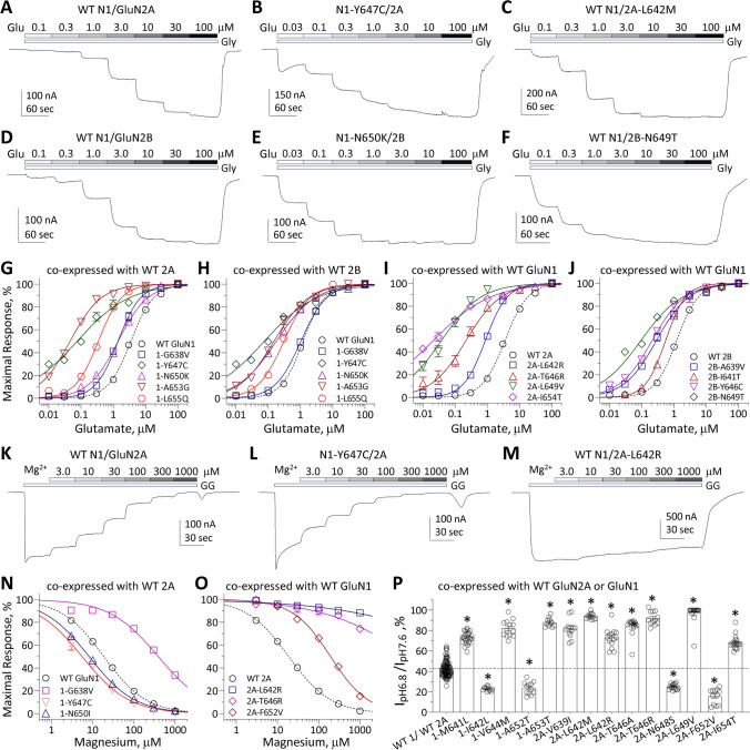
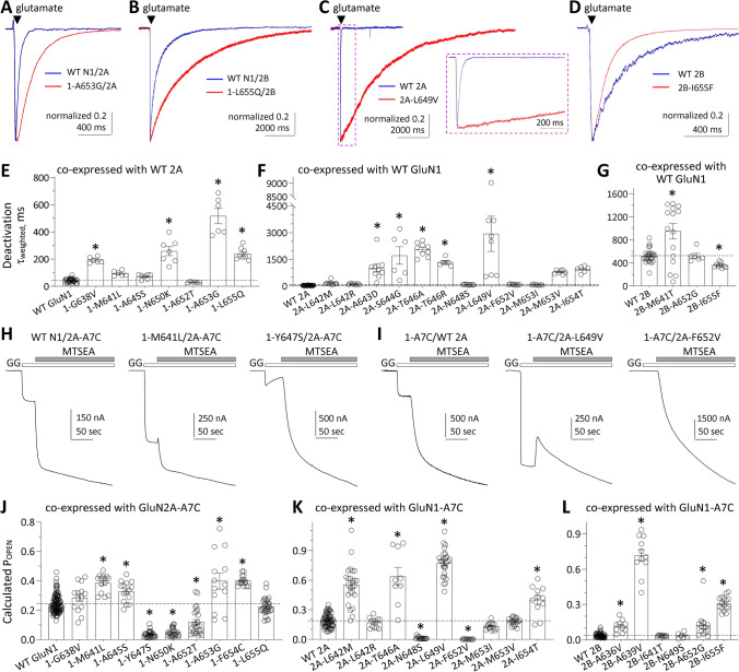
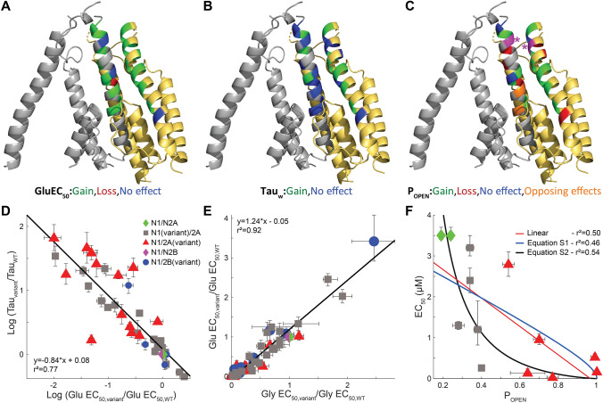
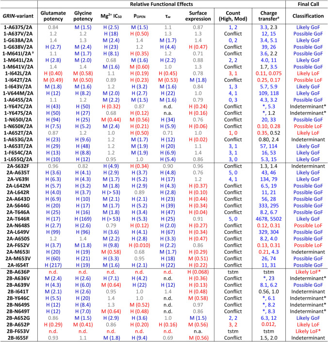
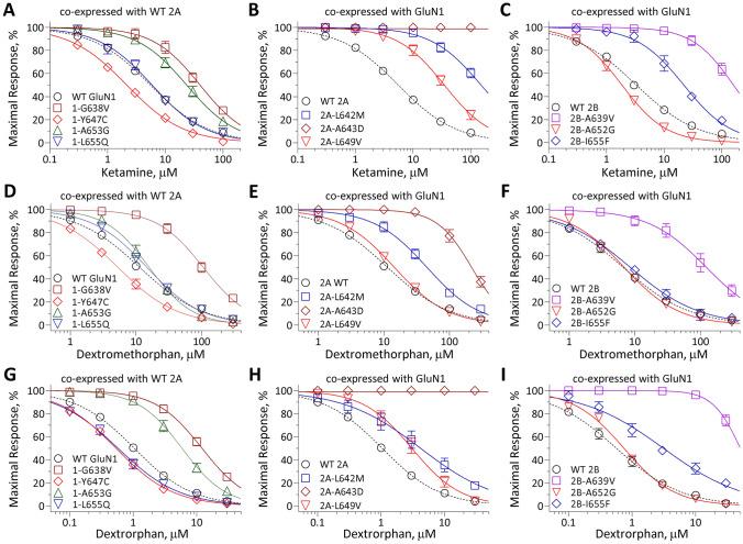
N-methyl-D-aspartate receptors (NMDARs) are members of the glutamate receptor family and participate in excitatory postsynaptic transmission throughout the central nervous system. Genetic variants in GRIN genes encoding NMDAR subunits are associated with a spectrum of neurological disorders. The M3 transmembrane helices of the NMDAR couple directly to the agonist-binding domains and form a helical bundle crossing in the closed receptors that occludes the pore. The M3 functions as a transduction element whose conformational change couples ligand binding to opening of an ion conducting pore. In this study, we report the functional consequences of 48 de novo missense variants in GRIN1, GRIN2A, and GRIN2B that alter residues in the M3 transmembrane helix. These de novo variants were identified in children with neurological and neuropsychiatric disorders including epilepsy, developmental delay, intellectual disability, hypotonia and attention deficit hyperactivity disorder. All 48 variants in M3 for which comprehensive testing was completed produce a gain-of-function (28/48) compared to loss-of-function (9/48); 11 variants had an indeterminant phenotype. This supports the idea that a key structural feature of the M3 gate exists to stabilize the closed state so that agonist binding can drive channel opening. Given that most M3 variants enhance channel gating, we assessed the potency of FDA-approved NMDAR channel blockers on these variant receptors. These data provide new insight into the structure-function relationship of the NMDAR gate, and suggest that variants within the M3 transmembrane helix produce a gain-of-function.

Keywords: Channelopathy; Functional genomics; Glutamate receptor; NMDA receptor; TMD.


Conflict of interest statement

SFT is a member of the SAB for Sage Therapeutics, Eumentis Therapeutics, the GRIN2B Foundation, the CureGRIN Foundation, and CombinedBrain. SFT is consultant for GRIN Therapeutics and Neurocrine, a cofounder of NeurOp, Inc. and Agrithera, and a member of the Board of Directors for NeurOp Inc. HY is the PI on a research grant from Sage Therapeutics to Emory and SJM is PI on a grant from GRIN Therapeutics to Emory. TAB—Consultancy for AveXis, Ovid, GW Pharmaceuticals, International Rett Syndrome Foundation, Takeda, Taysha, CureGRIN, GRIN Therapeutics, Alcyone, Neurogene, and Marinus; Clinical Trials with Acadia, Ovid, GW Pharmaceuticals, Marinus and RSRT; all remuneration has been made to his department.

Figures

Fig. 1
Fig. 1
Locations of disease-associated missense variants in the M3 transmembrane helix. A A linear schematic showing domain architecture of the GRIN/GluN and protein residue sequence alignment for highly conserved M3 transmembrane domain (TMD) across GluN1 and GluN2 subunits. NTD indicates the N-terminal domain (also known as ATD, amino terminal domain); S1 and S2 denote the first and second polypeptide sequences comprising the agonist binding domain (ABD). The aligned amino-acid sequence of the M3 helix of each NMDAR subunits is listed with a raster plot depicting the missense and synonymous variants found in gnomAD along with the 3DMTR score for these stretches, shown as a colorimetric raster plot. The red dashed box indicates the M3 transmembrane helix. B Homology model of the GluN1/GluN2A receptor built from previously reported GluN1/GluN2B cryo-EM data [20]. C The residues harboring missense variants are highlighted with different colors in a side view; the side of the subunit facing the pore is indicated
Fig. 2
Fig. 2
Variants in the M3 transmembrane helix influence pharmacological and biophysical properties of NMDARs. A–F Representative two electrode voltage clamp current recordings from Xenopus oocytes expressing GluN1/GluN2A and GluN1/GluN2B wild type or variant NMDAR subunits, as indicated. The glutamate concentration–response relationship was determined by co-applying increasing concentrations of glutamate with maximally effective concentration of glycine (30-100 µM). G–J Composite concentration–response curves for glutamate in the presence of 30-100 µM glycine fitted with the Hill equation (see Methods). K–M Representative voltage clamp current recordings show the Mg2+ concentration–response relationship for GluN1/GluN2A wild type and variants (as indicated) determined by co-applying increasing concentrations of Mg2+ with 100 µM glutamate and 100 µM glycine. N, O Composite concentration–response curves are shown for Mg2+ inhibition recorded at a holding potential of –60 mV for wild type and variant GluN1/GluN2A. P Summary of the effects of GluN1 and GluN2A variants on proton sensitivity, evaluated by the ratio of current response at pH 6.8 to pH 7.6 at a holding potential of − 40 mV
Fig. 3
Fig. 3
Variants in the M3 transmembrane helix change NMDAR biophysical properties. A-D Representative whole cell current responses recorded under voltage clamp illustrate the deactivation time course for GluN1/GluN2A, GluN1/GluN2B, GluN1-A653G/GluN2A, GluN1-L655Q/GluN2B, GluN1/GluN2A-L649V, GluN1/GluN2B-I655F NMDARs in response to brief 2–6 ms application and rapid removal of 1 mM glutamate with 100 µM glycine present in all solutions. Variant responses were normalized to the peak amplitude of wild type NMDAR responses. Each variant showed a prolonged deactivation time course compared to WT NMDARs. EG Summary of deactivation time course weighted tau for NMDAR variants. H, I Representative two electrode voltage clamp current recordings from Xenopus oocytes expressing GluN1/GluN2A wild type or variant NMDAR subunits (as indicated) showing current responses evoked by the application of 100 µM glutamate and 100 µM glycine followed by co-application of maximally effective glutamate and glycine plus 0.2 mM MTSEA (see Methods). N1-A7C indicates GluN1-A652C and 2A-A7C indicates GluN2A-A650C, which are covalently modified by MTSEA to lock the channel open. JL Summary of calculated channel open probability (POPEN) evaluated by the degree of MTSEA potentiation at a holding potential of − 40 mV for GluN1 (J), GluN2A (K) and GluN2B variants (L) (see Methods). Data are shown as mean ± SEM
Fig. 4
Fig. 4
Effects of variants on tau deactivation, glutamate EC50, glycine EC50, and open probability (POPEN). A Measured EC50 values for GluN1 and GluN2A variants that were significantly different than WT controls are mapped onto a GluN1/GluN2A homology model of two subunits of the transmembrane pore (GluN1-gray, GluN2 yellow) based on the non-active GluN1/GluN2B structure from Chou et al. [20]. Variants that increase open probability are green, variants that decrease open probability are red, those without an effect are blue, and positions that have multiple variants at the same residue with opposing significantly different effects are orange. Residues that had multiple tested variants which either produced no significant alterations or significant effects in one direction were colored according to observed significant effects. B Measured deactivation tau weighted values for GluN1 and GluN2A variants that were significantly different than WT controls are colored as in (A). C Measured open probability values for GluN1 and GluN2A variants that were significantly different than WT controls are colored as in (A). The site at which a Cys residue is substituted and modified by MTSEA on GluN1 (grey) and on GluN2A (yellow) are shown in magenta with an asterisk. D The logarithm (Log, base ten) of the ratio of variant to WT tauweighted is plotted against the Log of the ratio of variant to WT glutamate EC50. The slope was − 0.84, the intercept was − 0.08, and the value for R2 was 0.77. E The Log of the ratio of variant to WT glycine EC50 is plotted against the Log of the ratio of variant to WT glutamate EC50. The slope was 1.24, the intercept was -0.05, and the value for R2 was 0.92. F The measured glutamate EC50 value is plotted against Po for variants (GluN1 and GluN2A) that increase open probability compared to WT GluN1/GluN2A NMDARs (see Supplemental Fig. S3 for the full dataset). Three regressions are shown; linear fit (red, r2 = 0.50), equation S1 (blue, r2 = 0.46, see Supplemental Fig. S3), and equation S2 (black, r2 = 0.54, see Supplemental Fig. S3). Error bars depict SEM for Tau and E values and 99% confidence intervals for EC50 values
Fig. 5
Fig. 5
Assessment of M3 variant-mediated changes in parameters supporting GoF and LoF status. Glutamate and glycine potency ratios are given as WT/variant EC50 because EC50 is reciprocally related to potency. Mg2+ IC50, open probability (POPEN), weighted tau (τw), and surface expression fold effects are given as variant/WT. Two sets of the pooled τw data for same day controls were used to obtain WT parameter values for comparison to variants (see Supplemental Table S5). A high (H) or moderate (M) confidence (see Myers et al. [13]) for the change is indicated; red is LoF and blue is GoF. The number of high (H) and moderate (M) changes are given Count when there is no conflict in the direction of change for two parameters. Conflicting and subthreshold variants were re-classified as Possible GoF or Possible LoF if the fold change in the synaptic (left number) or non-synaptic (right number) of relative charge transfer ratio (Variant/WT) was > 2.5-fold or < 0.4-fold. aCalculation of relative for charge transfer (Variant/WT) was from Myers et al., [13]. n.d. indicates response was too small to measure (tstm). *Indicates that experiments to assess tau were run and currents recorded, but they were too small to allow reliable determination of tau and thus we cannot determine whether the variant alters overall function
Fig. 6
Fig. 6
Variants in the M3 transmembrane helix influence sensitivity to FDA-approved NMDAR channel blocker memantine. A-F Representative two electrode voltage clamp current recordings from Xenopus oocytes expressing GluN1/GluN2A and GluN1/GluN2B WT or variant NMDAR subunits, as indicated. The FDA-approved NMDAR channel blocker memantine concentration–response relationship was determined by co-applying increasing concentrations of memantine (Mem) with maximally effective concentrations of glutamate and glycine (100 µM) at a holding potential of − 40 mV. G-I Composite concentration–response curves of memantine were assessed by TEVC recordings of Xenopus oocytes for GluN1 (G), GluN2A (H) and GluN2B (I) variants in the presence of maximally effective concentrations of agonists (100 µM glutamate and 100 µM glycine) at a holding potential of − 40 mV. Data are shown as mean ± SEM
Fig. 7
Fig. 7
Variants in the M3 transmembrane helix influence sensitivity to FDA-approved NMDAR channel blockers ketamine, dextromethorphan and its metabolite dextrorphan. Composite concentration–response curves of FDA-approved NMDAR channel blockers ketamine (A–C), dextromethorphan (D–F) and its CYP2D6 metabolite dextrorphan (G–I) were assessed by TEVC recordings of Xenopus oocytes in the presence of maximally effective concentrations of agonists (100 µM glutamate and 100 µM glycine) at holding potential of – 40 mV. Data are shown as mean ± SEM

References

    1. Hansen KB, Wollmuth LP, Bowie D, Furukawa H, Menniti FS, Sobolevsky AI, Swanson GT, Swanger SA, Greger IH, Nakagawa T, McBain CJ, Jayaraman V, Low CM, Dell'Acqua ML, Diamond JS, Camp CR, Perszyk RE, Yuan H, Traynelis SF. Structure, function, and pharmacology of glutamate receptor ion channels. Pharmacol Rev. 2021;73(4):298–487. doi: 10.1124/pharmrev.120.000131. - DOI - PMC - PubMed
    1. Myers SJ, Yuan H, Kang JQ, Tan FCK, Traynelis SF, Low CM. Distinct roles of GRIN2A and GRIN2B variants in neurological conditions. F1000Res. 2019 doi: 10.12688/f1000research.18949.1. - DOI - PMC - PubMed
    1. Cenci MA, Skovgard K, Odin P. Non-dopaminergic approaches to the treatment of motor complications in Parkinson's disease. Neuropharmacology. 2022;210:109027. doi: 10.1016/j.neuropharm.2022.109027. - DOI - PubMed
    1. Geoffroy C, Paoletti P, Mony L. Positive allosteric modulation of NMDA receptors: mechanisms, physiological impact and therapeutic potential. J Physiol. 2022;600(2):233–259. doi: 10.1113/JP280875. - DOI - PubMed
    1. Li S, Stern AM. Bioactive human Alzheimer brain soluble Abeta: pathophysiology and therapeutic opportunities. Mol Psychiatry. 2022;27(8):3182–3191. doi: 10.1038/s41380-022-01589-5. - DOI - PubMed

Substances