. 2024 Apr;202(2):179-187.
doi: 10.1007/s00408-024-00672-8. Epub 2024 Mar 27.

Dysphagia as a Missing Link Between Post-surgical- and Opioid-Related Pneumonia

Affiliations

Dysphagia as a Missing Link Between Post-surgical- and Opioid-Related Pneumonia

Michael Frazure et al. Lung. 2024 Apr.

Abstract

Purpose: Postoperative pneumonia remains a common complication of surgery, despite increased attention. The purpose of our study was to determine the effects of routine surgery and post-surgical opioid administration on airway protection risk.

Methods: Eight healthy adult cats were evaluated to determine changes in airway protection status and for evidence of dysphagia in two experiments. (1) In four female cats, airway protection status was tracked following routine abdominal surgery (spay surgery) plus low-dose opioid administration (buprenorphine 0.015 mg/kg, IM, q8-12 h; n = 5). (2) Using a cross-over design, four naive cats (2 male, 2 female) were treated with moderate-dose (0.02 mg/kg) or high-dose (0.04 mg/kg) buprenorphine (IM, q8-12 h; n = 5).

Results: Airway protection was significantly affected in both experiments, but the most severe deficits occurred post-surgically as 75% of the animals exhibited silent aspiration.

Conclusion: Oropharyngeal swallow is impaired by the partial mu-opioid receptor agonist buprenorphine, most remarkably in the postoperative setting. These findings have implications for the prevention and management of aspiration pneumonia in vulnerable populations.

Keywords: Aspiration; Dysphagia; Opioid; Pneumonia; Postoperative; Swallow.


Conflict of interest statement

Disclosures: No conflicts of interest, financial or otherwise, are declared by the authors.

Figures

Figure 1.
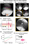
Figure 1.. Dysphagia following post-operative low-dose buprenorphine administration.
Swallow was evaluated using lateral plane videofluoroscopy. A) Anatomic orientation of the oropharynx, pharynx, cervical esophagus, larynx, and trachea. A penny, attached to the bowl, is included for calibration of measurements. Swallowing studies were obtained 48-hours after routine abdominal surgery (ovariohysterectomy), in voluntarily feeding cats 1-hour post intramuscular buprenorphine (0.015 mg/kg) administration. All phases of swallow were impacted. B) Three animals presented with significant aspiration without any overt signs or symptom (i.e., silent aspiration). Video 1 (yellow box) demonstrates a feeding bout with increasing amount of contrast agent traveling into the lower airways. C) The Airway Invasion Scale (AIS) is an ordinal scale, which documents the depth material traveled into the airway during swallow. During the control period all animals-maintained airway protection during purée and thin liquid trials, in contrast to post-surgery and opioid administration when penetration and/or aspiration was present in all animals. A Wilcoxon signed-rank test detected a significant increase in AIS scores (z = −2.21, p = 0.03). D) LSD post-hoc tests revealed that pharyngeal phase duration (onset of hyolaryngeal excursion through upper-esophageal sphincter (UES) closure and return of pharyngeal air space) was longer during post-operative purée feeding (424±28ms), compared to control (361±37ms), [F(2, 9)=8.1, p=0.01]. E) Maximum cervical esophageal distension before peristalsis was increased during post-operative purée feeding (12.4±0.9mm) compared to the control period (10.7± 0.9mm), [F(2, 9)=7.01, p=0.015]. Swallow durations are reported in ms. Distension measures are reported in mm. *p < 0.05.
Figure 2.
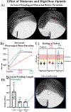
Figure 2.. Swallow efficiency is reduced after moderate and high dose opioid administration.
A) Representative videofluoroscopic image depicts increased maximum cervical esophageal distension after 48-hours on a lower-end clinical dose of buprenorphine (0.02 mg/kg), suggesting decreased sensitivity of esophageal distension reflexes. Maximum cervical esophageal distension before peristalsis increased on average following 48-hours on 0.02 mg/kg (13±2 mm) and 0.04 mg/kg buprenorphine (13±0.8 mm) compared to control (12±2 mm) but did not reach statistical significance. B) Pharyngeal swallow duration increased during thin liquid feeding after clinical doses of buprenorphine. Analysis of variance (ANOVA) showed differences in pharyngeal swallow duration in the 0.02 mg/kg [F(2, 8) = 4.62, p = 0.04] and 0.04 mg/kg [F(2, 7) = 27.6, p < 0.001] data sets. LSD post-hoc test results showed that pharyngeal swallow duration was longer after 48-hours on 0.02 mg/kg (322 ± 26ms) and 0.04 mg/kg buprenorphine (322 ± 13ms) compared to control (271±11ms). C) A Wilcoxon signed-rank test indicated a change in AIS ratings, with more scores indicative of airway invasion after 48-hours on 0.02 mg/kg buprenorphine (z = −2, p = 0.05). The increase in the ordinal AIS ratings describes that thin liquid laryngeal penetration occurred in all three animals that fed at this time point. AIS ratings were elevated in two of three animals after 48-hours on 0.04 mg/kg buprenorphine, but the effect was non-significant as a group. There was non-transient penetration to the vocal folds in one animal and small volume aspiration in one animal. D) Feeding bout length decreased during thin liquid feeding after buprenorphine administration. ANOVA showed differences in feeding length in the 0.04 mg/kg data set [F(2, 7) = 5.3, p=0.04]. LSD post-hoc test results showed that there were fewer swallows per bout after 24-hours (8±2) and 48-hours (7±1) on 0.04 mg/kg buprenorphine compared to control (21 ± 10). One animal demonstrated total feeding refusal at both time points after 0.04 mg/kg buprenorphine. E) Video 2 shows retrograde flow below the pharyngoesophageal segment, and trace aspiration during thin-liquid feeding in a female animal after 48-hours on 0.04 mg/kg buprenorphine. Of note: *p < 0.05, **p < 0.01, # indicates feeding refusal.

References

    1. Haller L, et al., Post-Operative Dysphagia in Anterior Cervical Discectomy and Fusion. Ann Otol Rhinol Laryngol, 2022. 131(3): p. 289–294. - PubMed
    1. Goeze A, et al., [Post-operative prevalence of dysphagia in head-and-neck cancer patients in the acute care units]. Laryngorhinootologie, 2022. 101(4): p. 320–326. - PubMed
    1. Greenberg JA, et al., Evaluation of post-operative dysphagia following anti-reflux surgery. Surg Endosc, 2022. 36(7): p. 5456–5466. - PubMed
    1. Hayes M, et al., Prevalence, nature and trajectory of dysphagia postoesophageal cancer surgery: a prospective longitudinal study protocol. BMJ Open, 2022. 12(9): p. e058815. - PMC - PubMed
    1. Nath A, et al., Dysphagia after vertical sleeve gastrectomy: Evaluation of risk factors and assessment of endoscopic intervention. World J Gastroenterol, 2016. 22(47): p. 10371–10379. - PMC - PubMed

Publication types