. 2024 Apr 29;199(1):49-62.
doi: 10.1093/toxsci/kfae030.

A whale of a tale: whale cells evade the driving mechanism for hexavalent chromium-induced chromosome instability

Affiliations

A whale of a tale: whale cells evade the driving mechanism for hexavalent chromium-induced chromosome instability

Haiyan Lu et al. Toxicol Sci. .

Abstract

Chromosome instability, a hallmark of lung cancer, is a driving mechanism for hexavalent chromium [Cr(VI)] carcinogenesis in humans. Cr(VI) induces structural and numerical chromosome instability in human lung cells by inducing DNA double-strand breaks and inhibiting homologous recombination repair and causing spindle assembly checkpoint (SAC) bypass and centrosome amplification. Great whales are long-lived species with long-term exposures to Cr(VI) and accumulate Cr in their tissue, but exhibit a low incidence of cancer. Data show Cr(VI) induces fewer chromosome aberrations in whale cells after acute Cr(VI) exposure suggesting whale cells can evade Cr(VI)-induced chromosome instability. However, it is unknown if whales can evade Cr(VI)-induced chromosome instability. Thus, we tested the hypothesis that whale cells resist Cr(VI)-induced loss of homologous recombination repair activity and increased SAC bypass and centrosome amplification. We found Cr(VI) induces similar amounts of DNA double-strand breaks after acute (24 h) and prolonged (120 h) exposures in whale lung cells, but does not inhibit homologous recombination repair, SAC bypass, or centrosome amplification, and does not induce chromosome instability. These data indicate whale lung cells resist Cr(VI)-induced chromosome instability, the major driver for Cr(VI) carcinogenesis at a cellular level, consistent with observations that whales are resistant to cancer.

Keywords: DNA double-strand break; chromosome instability; hexavalent chromium; homologous recombination repair; whale.


Figures

Figure 1.
Figure 1.
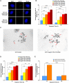
Particulate Cr(VI) induces DNA double-strand breaks in bowhead whale lung cells. Data reflect the mean of at least 3 independent experiments. Error bars = Standard error of the mean. *Significantly different from untreated cells at the same timepoint (#p <.1, *p <.05, **p <.01, ***p <.001, ****p <.0001). A, Representative images of γ-H2AX foci in bowhead whale lung cells. B, Breaks measured as γ-H2AX foci. C, Breaks measured with the neutral comet assay. D, Breaks measured in bowhead whale compared with human lung cells measured with the neutral comet assay after 120 h particulate Cr(VI) exposure. Human data originally published as tail moment in Qin et al. (2014), reanalyzed here as tail intensity.
Figure 2.
Figure 2.
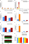
Particulate Cr(VI) induces cell cycle arrest in bowhead whale lung cells. Data reflect the mean of at least 3 independent experiments. Error bars = Standard error of the mean. *Significantly different from untreated cells at the same timepoint (*p <.05, **p <.01). (A) and (B) are the flow cytometry histograms of cell cycle profiles. A, Cell cycle profiles after 24 h zinc chromate. B, Cell cycle profiles after 120 h zinc chromate exposure. C, Flow cytometry gating analysis after 24 h zinc chromate exposure. D, Flow cytometry gating analysis after 120 h exposure. E, Mitotic index after 24 or 120 h zinc chromate exposure in bowhead whale lung cells.
Figure 3.
Figure 3.
Particulate Cr(VI) induced-DNA double-strand breaks occur in the S and G2/M phases in bowhead whale lung cells and human lung cells. Analysis of γ-H2AX in cell cycle phase by flow cytometry. Data reflect the mean of at least 3 independent experiments. Error bars = standard error of the mean. *Significantly different from untreated cells at the same timepoint (*p <.05, **p <.01). A, 24 h exposure in bowhead lung cells. B, 120 h exposure in bowhead lung cells. C, 24 h exposure in human lung cells. D, 120 h exposure in human lung cells.
Figure 4.
Figure 4.
Particulate Cr(VI) induces homologous recombination repair activity in bowhead whale lung cells. Data represent an average of 3 experiments. Error bars = standard error of the mean. *Significantly different from untreated cells at the same timepoint (*p <.05, **p <.01, ***p <.001, ****p <.0001). & data from 1 experiment, 2 experiments produced no metaphases. @ data from 3 experiments but 1 experiment only had 28 metaphases. A, Representative images of RAD51 foci bowhead lung cells. B, RAD51 foci formation. C, Representative images of SCEs, indicated by red arrows, in bowhead lung cells. D, SCE formation in bowhead lung cells. E, SCE formation in human lung cells.
Figure 5.
Figure 5.
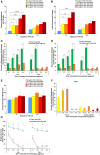
Cr(VI) does not alter securin levels and does not induce spindle assembly checkpoint (SAC) bypass or centrosome amplification in whale cells. Human data are added here for ease of comparison and were originally reported in Holmes et al. (2010). A, SAC bypass shown as the sum of stacked columns of centromere spreading, premature centromere division, and premature anaphase. Data reflect the mean of 3 independent experiments +/− the standard error of the mean. B, SAC bypass shows as the sum of stacked columns of centromere spreading, premature centromere division, and premature anaphase in bowhead whale lung cells compared with human lung cells. Data for bowhead whale lung is the same as panel A, graphed here on a scale that fits the human data. NM = not enough metaphases to analyze treatment. Data reflect the mean of 3 independent experiments +/− the standard error of the mean. C, Centrosome amplification in interphase cells. Data reflect the mean of 2 independent experiments. D, Centrosome amplification in interphase cells in bowhead whale lung cells compared with human lung cells. Data for bowhead whale lung is the same as panel C, graphed here on a scale that fits the human data. Human data reflect the mean of 3 independent experiments +/− the standard error of the mean. E, Centrosome amplification in mitotic cells. Data reflect the mean of 2 independent experiments. F, Centrosome amplification in mitotic cells in bowhead whale lung cells compared with human lung cells. Data for bowhead whale lung is the same as panel E, graphed here on a scale that fits the human data. Human data reflect the mean of 3 independent experiments +/− the standard error of the mean. G, Representative Western blot for securin. α-tubulin was used as a loading control. H, Securin whole cell protein levels. Data are expressed as percent of untreated control cells and reflect the mean of 3 independent experiments +/− the standard error of the mean.
Figure 6.
Figure 6.
Cr(VI) does not induce structural or numerical chromosome instability and is less genotoxic and cytotoxic in whale lung cells. Data represent an average of at least 3 experiments. Error bars = standard error of the mean. The human chromosome instability data in this figure (panels C–F) are added here for ease of comparison and were originally reported in Holmes et al. (2010). *Significantly different from untreated cells at the same timepoint (#p <.1, **p <.01, ***p <.001, ****p <.0001). NM = not enough metaphases to analyze treatment. A, Structural chromosome instability measured as percent of metaphases with damage. B, Structural chromosome instability measured as total chromosome aberrations in 100 metaphases. C, Structural chromosome instability measured as percentage of metaphases with damage in bowhead whale lung cells compared with human lung cells. Data for bowhead whale lung is the same as panel A, graphed here on a scale that fits the human data. D, Structural chromosome instability measured as total chromosome aberrations in 100 metaphases in bowhead whale lung cells compared with human lung cells. Data for bowhead whale lung is the same as panel B, graphed here on a scale that fits the human data. E, Numerical chromosome instability measured as percent of metaphases with aneuploidy. F, Numerical chromosome instability measured as percent of metaphases with aneuploidy in bowhead whale lung cells compared with human lung cells. Data for bowhead whale lung is the same as panel E, graphed here on a scale that fits the human data. G, Cytotoxicity in bowhead whale cells compared with human lung cells. The significant differences between bowhead whale lung cells and human lung cells at 0.1, 0.2, 0.3, and 0.4 after 24 h exposure are ap < .01, bp < .0001, cp < .0001, and dp < .0001. The significant differences between bowhead whale lung cells and human lung cells at 0.1, 0.2, 0.3, and 0.4 after 120 h exposure are ep < .0001, fp < .0001, gp < .0001, and hp < .0001.

References

    1. Blasiak J. (2021). Single-strand annealing in cancer. IJMS. 22, 2167. - PMC - PubMed
    1. Brinkley B. R. (2001). Managing the centrosome numbers game: From chaos to stability in cancer cell division. Trends Cell Biol. 11, 18–21. - PubMed
    1. Browning C. L., Qin Q., Kelly D. F., Prakash R., Vanoli F., Jasin M., Wise J. P. Sr (2016). Prolonged particulate hexavalent chromium exposure suppresses homologous recombination repair in human lung cells. Toxicol. Sci. 153, 70–78. - PMC - PubMed
    1. Browning C. L., Wise C. F., Wise J. P. Sr (2017). Prolonged particulate chromate exposure does not inhibit homologous recombination repair in North Atlantic right whale (Eubalaena glacialis) lung cells. Toxicol. Appl. Pharmacol. 331, 101–107. - PMC - PubMed
    1. Bryant H. E., Ying S., Helleday T. (2006). Homologous recombination is involved in repair of chromium-induced DNA damage in mammalian cells. Mutat. Res. 599, 116–123. - PubMed

Publication types

MeSH terms

LinkOut - more resources