Review
. 2024 Feb 29;11(3):290.
doi: 10.3390/children11030290.

Image-Guided Percutaneous Drainage of Abdominal Abscesses in Pediatric Patients

Affiliations
Review

Image-Guided Percutaneous Drainage of Abdominal Abscesses in Pediatric Patients

Dimitrij Kuhelj et al. Children (Basel). .

Abstract

Image-guided percutaneous abscess drainage (IPAD) is an effective, minimally invasive technique to manage infected abdominal fluid collections in children. It is the treatment of choice in cases where surgery is not immediately required due to another coexisting indication. The skills and equipment needed for this procedure are widely available. IPAD is typically guided by ultrasound, fluoroscopy, computed tomography, or a combination thereof. Abscesses in hard-to-reach locations can be drained by intercostal, transhepatic, transgluteal, transrectal, or transvaginal approaches. Pediatric IPAD has a success rate of over 80% and a low complication rate.

Keywords: Seldinger technique; appendicitis; children; pediatrics; percutaneous abscess drainage.


Conflict of interest statement

The authors declare no conflicts of interest.

Figures

Figure 1
Figure 1
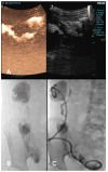
T (A) An ultrasound image showing an encapsulated fluid collection in the right hemiabdomen. (B) The caudal-most part of the multiloculated collection extended retrovesically. (C,D) Fluoroscopic image of image-guided percutaneous abscess drainage (IPAD) using a 10F drainage catheter.
Figure 2
Figure 2
(A) One week after the IPAD, no clinical improvement was observed in the patient, and a follow-up contrast-enhanced ultrasound was performed injecting the SonoVue® contrast directly into the abscess cavity using the inserted drainage catheter. CEUS revealed the communication between the iliac and paracolic abscess. (B) Another IPAD was immediately performed. The iodine contrast applied during fluoroscopy revealed three communicating abscess cavities that filled with the contrast applied under pressure, but the paracolic cavity then failed to drain upon iliac cavity drainage. (C) Then, a separate paracolic 10F catheter was inserted in addition to another new catheter, placed in the retrovesical abscess cavity. After ten days, the US follow-up revealed no signs of abscess (not depicted), and the patient was discharged.

References

    1. Wallace M.J., Chin K.W., Fletcher T.B., Bakal C.W., Cardella J.F., Grassi C.J., Grizzard J.D., Kaye A.D., Kushner D.C., Larson P.A., et al. Quality Improvement Guidelines for Percutaneous Drainage/Aspiration of Abscess and Fluid Collections. J. Vasc. Interv. Radiol. 2010;21:431–435. doi: 10.1016/j.jvir.2009.12.398. - DOI - PubMed
    1. Gervais D.A., Brown S.D., Connolly S.A., Brec S.L., Harisinghani M.G., Mueller P.R. Percutaneous Imaging-Guided Abdominal and Pelvic Abscess Drainage in Children. RadioGraphics. 2004;24:737–754. doi: 10.1148/rg.243035107. - DOI - PubMed
    1. McGahan J.P. The History of Interventional Ultrasound. J. Ultrasound Med. 2004;23:727–741. doi: 10.7863/jum.2004.23.6.727. - DOI - PubMed
    1. van Sonnenberg E., Wittich G.R., Edwards D.K., Casola G., von Waldenburg Hilton S., Self T.W., Keightley A., Withers C. Percutaneous Diagnostic and Therapeutic Interventional Radiologic Procedures in Children: Experience in 100 Patients. Radiology. 1987;162:601–605. doi: 10.1148/radiology.162.3.2949336. - DOI - PubMed
    1. Newman K., Ponsky T., Kittle K., Dyk L., Throop C., Gieseker K., Sills M., Gilbert J. Appendicitis 2000: Variability in Practice, Outcomes, and Resource Utilization at Thirty Pediatric Hospitals. J. Pediatr. Surg. 2003;38:372–379. doi: 10.1053/jpsu.2003.50111. - DOI - PubMed

LinkOut - more resources