. 2024 Mar 17;16(6):1180.
doi: 10.3390/cancers16061180.

PIP4K2B Protein Regulation by NSD1 in HPV-Negative Head and Neck Squamous Cell Carcinoma

Affiliations

PIP4K2B Protein Regulation by NSD1 in HPV-Negative Head and Neck Squamous Cell Carcinoma

Iuliia Topchu et al. Cancers (Basel). .

Abstract

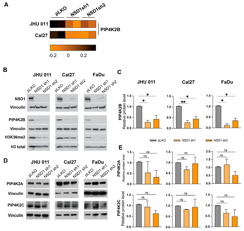
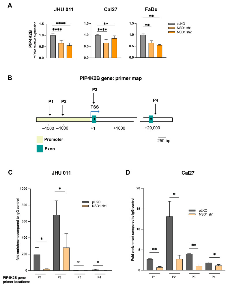
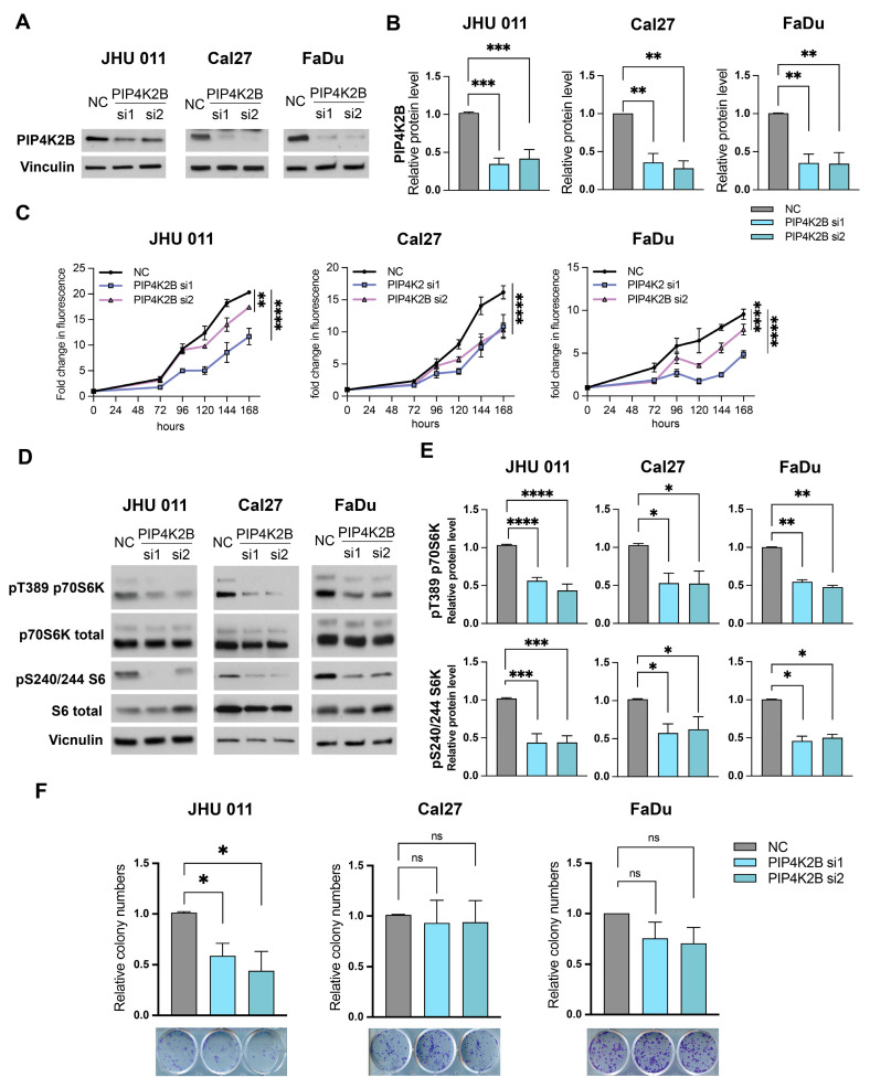
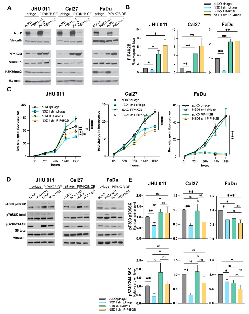
Head and neck squamous cell carcinoma (HNSCC) ranks among the most prevalent global cancers. Despite advancements in treatments, the five-year survival rate remains at approximately 66%. The histone methyltransferase NSD1, known for its role in catalyzing histone H3 lysine 36 di-methylation (H3K36me2), emerges as a potential oncogenic factor in HNSCC. Our study, employing Reverse Phase Protein Array (RPPA) analysis and subsequent validation, reveals that PIP4K2B is a key downstream target of NSD1. Notably, PIP4K2B depletion in HNSCC induces downregulation of the mTOR pathway, resulting in diminished cell growth in vitro. Our investigation highlights a direct, positive regulatory role of NSD1 on PIP4K2B gene transcription through an H3K36me2-dependent mechanism. Importantly, the impact of PIP4K2B appears to be context-dependent, with overexpression rescuing cell growth in laryngeal HNSCC cells but not in tongue/hypopharynx cells. In conclusion, our findings implicate PIP4K2B as a novel NSD1-dependent protein in HNSCC, suggesting its potential significance for laryngeal cancer cell survival. This insight contributes to our understanding of the molecular landscape in HNSCC and establishes PIP4KB as a promising target for drug development.

Keywords: NSD1; PIP4K; PIP4K2B; head and neck cancer squamous cell carcinoma (HNSCC); mTORC1.


Conflict of interest statement

All authors declare no conflicts of interest for this study.

Figures

Figure 1
Figure 1
NSD1 regulates PIK4K2B protein, but not other PIP4K family members’ expression in head and neck cancer cell lines. (A) Heatmap of the RPPA results for PIP4K2B protein in JHU 011 and Cal27 cell lines. (B) Representative images of validation of the PIP4K2B protein level by Western blot in JHU 011, Cal27, and FaDu cell lines with NSD1 knockdown after 72 h doxycycline induction. (C) Quantification of Western blot images in (B). Statistical significance is determined by the Mann–Whitney test, pairwise comparing every knockdown (sh1 and sh2) with control (pLKO). (D) Representative Western blot images of the PIP4K2A and PIP4K2C protein levels in JHU 011, Cal27, and FaDu cell lines with NSD1 knockdown after 72h doxycycline induction. (E) Quantification of Western blot images in (D). Statistical significance is determined by the Mann–Whitney test, pairwise comparing every knockdown (sh1 and sh2) with control (pLKO). Data are performed from at least three independent biological repeats. The error bars are presented as mean ± SEM. ns—not significant, * p < 0.05, ** p < 0.01.
Figure 2
Figure 2
NSD1 regulates PIP4K2B in a direct manner. (A) mRNA level of the PIP4K2B gene in JHU 011, Cal27, and FaDu cell lines with NSD1 knockdown after 72 h of doxycycline induction measured by qRT-PCR. PIP4K2B relative level was normalized on 18S as a control gene. Statistical significance is determined by the Mann–Whitney test, pairwise comparing every knockdown (sh1 and sh2) with control (pLKO). (B) Primer location map of the PIP4K2B gene regions for ChIP-qPCR. TSS—transcription start site; P—primers. (C) ChIP-qPCR with H3K36me2 antibodies on the PIP4K2B gene in JHU 011 cell line with NSD1 knockdown after 72 h doxycycline induction. Statistical significance is determined by the Mann–Whitney test, pairwise comparing every knockdown (sh1) with control (pLKO). (D) ChIP-qPCR with H3K36me2 antibodies on the PIP4K2B gene in Cal27 cell line with NSD1 knockdown after 72 h doxycycline induction. Statistical significance is determined by the Mann–Whitney test, pairwise comparing every knockdown (sh1) with control (pLKO). Data are performed from at least three independent biological repeats. The error bars are presented as mean ± SEM. ns—not significant, * p < 0.05, ** p < 0.01, **** p < 0.0001.
Figure 3
Figure 3
Depletion of PIP4K2B results in reduced cell growth and diminished mTOR signaling in head and neck cancer cells. (A) Representative images of validation of the PIP4K2B protein level by Western blot in JHU 011, Cal27, and FaDu cell lines with PIP4K2B siRNA knockdown after 72 h transfection. (B) Quantification of Western blot images in (A). Statistical significance is determined by the Mann–Whitney test, pairwise comparing every knockdown (si1 and si2) with negative control (NC). (C) Proliferation JHU 011, Cal27, and FaDu cell lines transfected with siRNA against PIP4K2B, as measured by CTB assay for up to 168 h, at indicated time points. Statistical significance was determined by ANOVA with Dunnett multiple comparison post-test. Each group was compared to the negative control (NC). (D) Representative Western blot images of p70S6K and S6 protein levels in phospho- and total isoforms in JHU 011, Cal27, FaDu cell lines with PIP4K2B siRNA knockdown after 72 h transfection. (E) Quantification of Western blot images in (A). Statistical significance is determined by the Mann–Whitney test, pairwise comparing every knockdown (si1 and si2) with negative control (NC). (F) Representative images of colony formation assay and quantification of the relative colony numbers in JHU 011, Cal27, FaDu cell lines with PIP4K2B siRNA knockdown. Statistical significance is determined by the Mann–Whitney test, pairwise comparing every knockdown (si1 and si2) with negative control (NC). Data are performed from at least three independent biological repeats. The error bars are presented as mean ± SEM. ns—not significant, * p < 0.05, ** p < 0.01, *** p < 0.001, and **** p < 0.0001.
Figure 4
Figure 4
PIP4K2B overexpression leads to cell proliferation restoration only in laryngeal cell line. (A) Representative images of validation of the PIP4K2B protein overexpression (PIP4K2B; pHage is an empty vector) by Western blot in JHU 011, Cal27, and FaDu cell lines with NSD1 knockdown after 72 h doxycycline induction. (B) Quantification of Western blot images in (A). Statistical significance is determined by the Mann–Whitney test, pairwise comparing as it is noted on the graph. (C) Proliferation JHU 011, Cal27, and FaDu cell lines with PIP4K2B overexpression protein along with NSD1 knockdown, as measured by CTB assay for up to 168 h, at indicated time points. Statistical significance was determined by ANOVA with Dunnett multiple comparison post-test. Each group was compared to the other. (D) Representative Western blot images of p70S6K and S6 protein levels in phospho- and total isoforms in JHU 011, Cal27, FaDu cell lines with PIP4K2B overexpression and NSD1 knockdown after 72 h doxycycline induction. (E) Quantification of Western blot images in (D). Statistical significance is determined by the Mann–Whitney test, pairwise comparing every group to other. Data are performed from at least three independent biological repeats. The error bars are presented as mean ± SEM. Ns—not significant, * p < 0.05, ** p < 0.01, *** p < 0.001, and **** p < 0.0001.

References

    1. Topchu I., Pangeni R.P., Bychkov I., Miller S.A., Izumchenko E., Yu J., Golemis E., Karanicolas J., Boumber Y. The role of NSD1, NSD2, and NSD3 histone methyltransferases in solid tumors. Cell Mol. Life Sci. 2022;79:285. doi: 10.1007/s00018-022-04321-2. - DOI - PMC - PubMed
    1. Husmann D., Gozani O. Histone lysine methyltransferases in biology and disease. Nat. Struct. Mol. Biol. 2019;26:880–889. doi: 10.1038/s41594-019-0298-7. - DOI - PMC - PubMed
    1. Lu T., Jackson M.W., Wang B., Yang M., Chance M.R., Miyagi M., Gudkov A.V., Stark G.R. Regulation of NF-kappaB by NSD1/FBXL11-dependent reversible lysine methylation of p65. Proc. Natl. Acad. Sci. USA. 2010;107:46–51. doi: 10.1073/pnas.0912493107. - DOI - PMC - PubMed
    1. Zhang S., Zhang F., Chen Q., Wan C., Xiong J., Xu J. CRISPR/Cas9-mediated knockout of NSD1 suppresses the hepatocellular carcinoma development via the NSD1/H3/Wnt10b signaling pathway. J. Exp. Clin. Cancer Res. 2019;38:1–14. doi: 10.1186/s13046-019-1462-y. - DOI - PMC - PubMed
    1. Chen Y., Li X., Xu J., Xiao H., Tang C., Liang W., Zhu X., Fang Y., Wang H., Shi J. Knockdown of nuclear receptor binding SET domain-containing protein 1 (NSD1) inhibits proliferation and facilitates apoptosis in paclitaxel-resistant breast cancer cells via inactivating the Wnt/β-catenin signaling pathway. Bioengineered. 2022;13:3526–3536. doi: 10.1080/21655979.2021.2018973. - DOI - PMC - PubMed

LinkOut - more resources