Review
. 2024 Mar 1;13(3):312.
doi: 10.3390/antiox13030312.

Reactive Oxygen Species Signaling and Oxidative Stress: Transcriptional Regulation and Evolution

Affiliations
Review

Reactive Oxygen Species Signaling and Oxidative Stress: Transcriptional Regulation and Evolution

Yuhang Hong et al. Antioxidants (Basel). .

Abstract

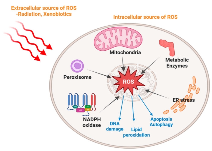
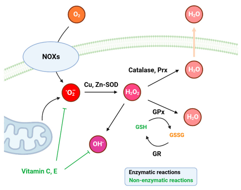
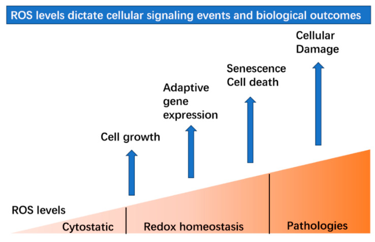
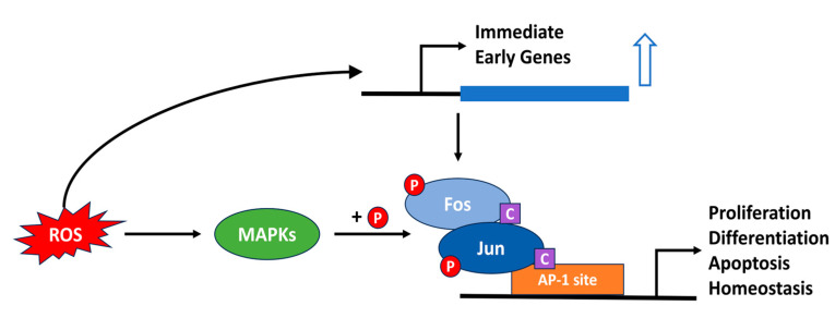
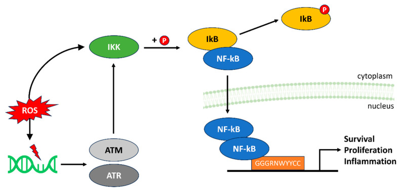
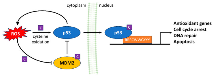
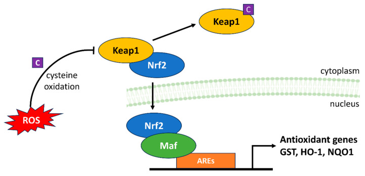
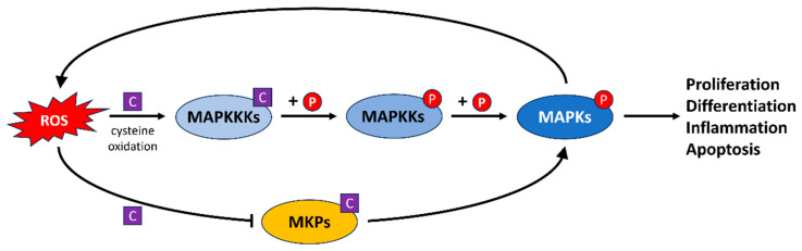
Since the evolution of the aerobic metabolism, reactive oxygen species (ROS) have represented significant challenges to diverse life forms. In recent decades, increasing knowledge has revealed a dual role for ROS in cell physiology, showing they serve as a major source of cellular damage while also functioning as important signaling molecules in various biological processes. Our understanding of ROS homeostasis and ROS-mediated cellular signaling pathways has presumed that they are ancient and highly conserved mechanisms shared by most organisms. However, emerging evidence highlights the complexity and plasticity of ROS signaling, particularly in animals that have evolved in extreme environments. In this review, we focus on ROS generation, antioxidative systems and the main signaling pathways that are influenced by ROS. In addition, we discuss ROS's responsive transcription regulation and how it may have been shaped over the course of evolution.

Keywords: DNA repair; cellular signaling; reactive oxygen species; transcriptional regulation; vertebrate evolution.


Conflict of interest statement

The authors declare no conflicts of interest.

Figures

Figure 1
Figure 1
Schematic representation of oxidative stress induced by intracellular and extracellular stimuli. Extracellular sources of ROS include environmental factors such as radiation and xenobiotics. Intracellular ROS are generated mainly in the mitochondria, as well as other organelles such as the peroxisome and endoplasmic reticulum, and in addition by some metabolic enzymes. Excessive ROS production can lead to cell apoptosis, autophagy, lipid peroxidation and DNA damage.
Figure 2
Figure 2
Schematic presentation of antioxidation systems with enzymatic and non-enzymatic antioxidants. Black arrows indicate enzymatic reactions and green lines indicate non-enzymatic reactions. NOXs: the NADPH oxidases; SOD: superoxide dismutase; Prx: peroxiredoxin; GPx: glutathione peroxidase; GR: glutathione reductase; GSH: glutathione; GSSG: glutathione disulfide.
Figure 3
Figure 3
ROS levels dictate different signaling events and biological outcomes. Within the range of normal physiological levels, ROS participate in many important cellular processes to maintain redox homeostasis. At low levels, ROS promote cell growth by proliferation and differentiation. The induction of ROS levels leads to adaptive gene expression such as the up-regulated expression of antioxidative genes. Then, exposure to higher levels will result in the initiation of senescence or cell death.
Figure 4
Figure 4
ROS-mediated regulation of the AP-1 transcription factor family. ROS act as signaling molecules, activating AP-1 protein complexes and enhancing their transcriptional activation function. A key step in this process is the phosphorylation of the AP-1 factors by MAPKs (+P). In addition, changes in the redox state of certain conserved cysteine residues (C) have been implicated in regulating DNA binding. Subsequently, the expression of genes involved in fundamental cellular processes is regulated. Excessive amounts of ROS lead to aberrant AP-1 activation, potentially disrupting cellular homeostasis.
Figure 5
Figure 5
ROS modulation of the NF-κB pathway. At moderate levels, ROS facilitate NF-κB activation and translocation to the nucleus to initiate gene transcription. This involves the phosphorylation of IkB by IKK (indicated by P), thereby targeting it for ubiquitin-mediated degradation and liberating NF-kB dimers, which translocate to the nucleus. Excessive ROS levels directly and indirectly disrupt IKK regulation, resulting in sustained activation of NF-κB and inflammatory responses.
Figure 6
Figure 6
ROS regulation of the p53 signaling pathway. ROS serve as both triggers and regulators of p53 activation in response to cellular stressors. At physiological concentrations, ROS stabilize and activate p53, promoting DNA repair and antioxidant responses, preventing spreading of damaged cells and keeping ROS at non-toxic levels. These effects of ROS on p53 as well as its regulatory factor MDM2 are mediated by oxidation of cysteine residues in both proteins (C). However, high p53 levels lead to pro-oxidant responses, increasing ROS and suppressing antioxidant genes.
Figure 7
Figure 7
ROS modulation of the Keap1-Nrf2-ARE signaling pathway. ROS play a central role in the balance between Nrf2 activation and inhibition. At physiological levels, Keap1 maintains Nrf2 at low concentrations. However, at high ROS concentrations, Keap1 is oxidized at particular cysteine residues (C) and Nrf2 degradation is prevented, thereby increasing transcription of antioxidant genes via AREs and promoting defense against oxidative stress.
Figure 8
Figure 8
ROS regulation of the MAPK signaling pathway. ROS serve as crucial regulators of the MAPK signaling pathway both directly by activating upstream kinases, and so enabling them to phosphorylate their downstream targets (P), and indirectly by inhibiting MAPK phosphatases via the oxidation of cysteine residues (C) on these enzymes. MAPKs can increase ROS levels, reinforcing the signal and its downstream effects.
Figure 9
Figure 9
Schematic representation of the ROS response of the D-box element in zebrafish and cavefish. In zebrafish cells (left panel), elevated levels of ROS are induced by exposure to both visible and UV light. Then, ROS activate MAPKs including p38 and JNK rapidly by phosphorylation and in turn activate the PAR factors. PAR factors bind to D-box enhancer elements in the promoters of a set of clock and DNA repair genes and ultimately lead to induced gene transcription and clock entrainment. In cavefish cells (right panel), this signaling fails to activate transcription of the same genes via the D-box [95].

References

    1. Bedard K., Krause K.-H. The NOX Family of ROS-Generating NADPH Oxidases: Physiology and Pathophysiology. Physiol. Rev. 2007;87:245–313. doi: 10.1152/physrev.00044.2005. - DOI - PubMed
    1. D’Autréaux B., Toledano M.B. ROS as signalling molecules: Mechanisms that generate specificity in ROS homeostasis. Nat. Rev. Mol. Cell Biol. 2007;8:813–824. doi: 10.1038/nrm2256. - DOI - PubMed
    1. Ray P.D., Huang B.-W., Tsuji Y. Reactive oxygen species (ROS) homeostasis and redox regulation in cellular signaling. Cell. Signal. 2012;24:981–990. doi: 10.1016/j.cellsig.2012.01.008. - DOI - PMC - PubMed
    1. Zhang J., Wang X., Vikash V., Ye Q., Wu D., Liu Y., Dong W. ROS and ROS-Mediated Cellular Signaling. Oxidative Med. Cell. Longev. 2016;2016:4350965. doi: 10.1155/2016/4350965. - DOI - PMC - PubMed
    1. Boveris A., Chance B. The mitochondrial generation of hydrogen peroxide. General properties and effect of hyperbaric oxygen. Biochem. J. 1973;134:707–716. doi: 10.1042/bj1340707. - DOI - PMC - PubMed

LinkOut - more resources