

Bioactive Lignan Honokiol Alleviates Ovarian Oxidative Stress in Aging Laying Chickens by Regulating SIRT3/AMPK Pathway
- PMID: 38539910
- PMCID: PMC10967992
- DOI: 10.3390/antiox13030377
Bioactive Lignan Honokiol Alleviates Ovarian Oxidative Stress in Aging Laying Chickens by Regulating SIRT3/AMPK Pathway
Abstract
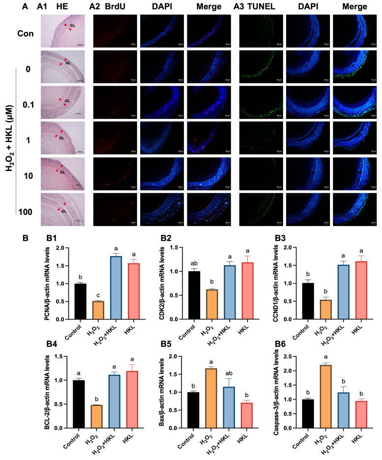
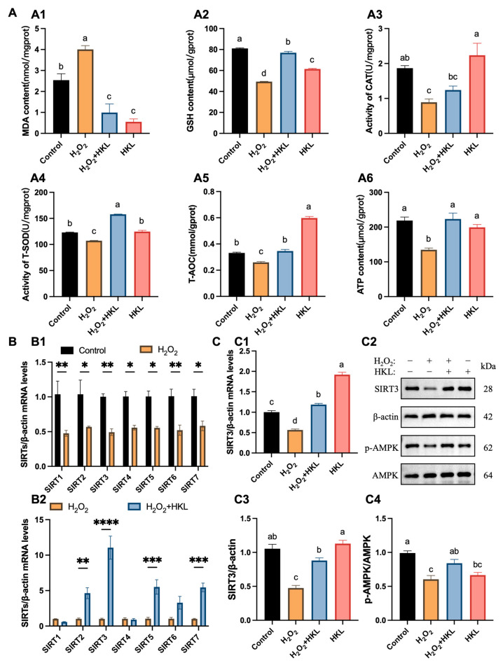
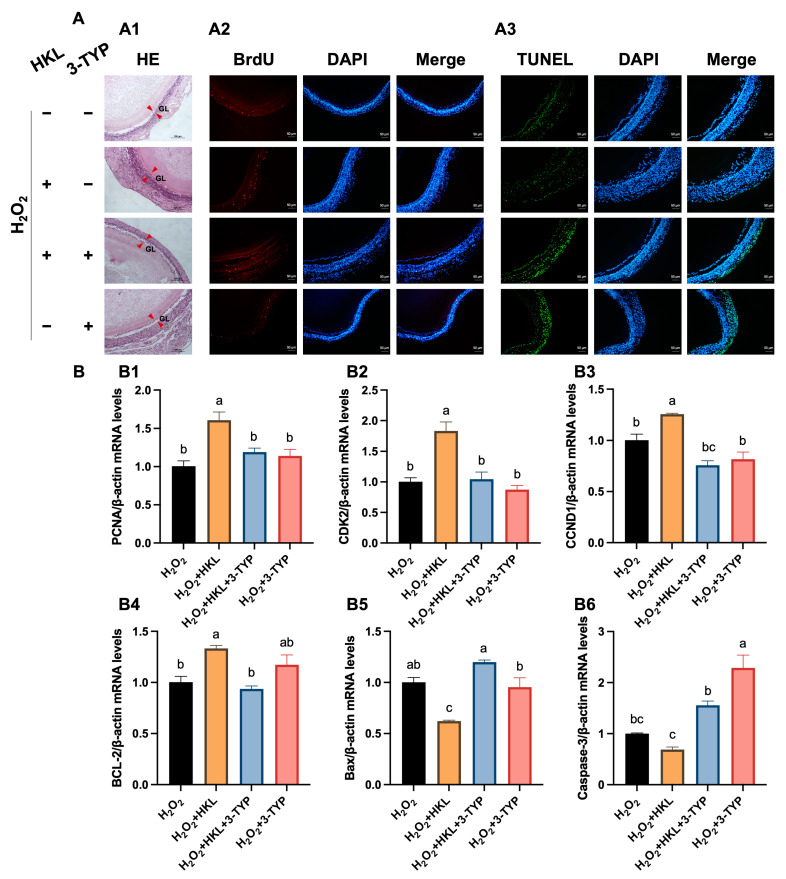
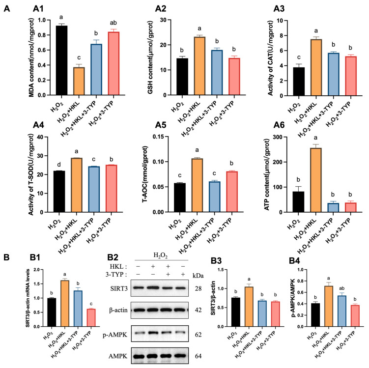
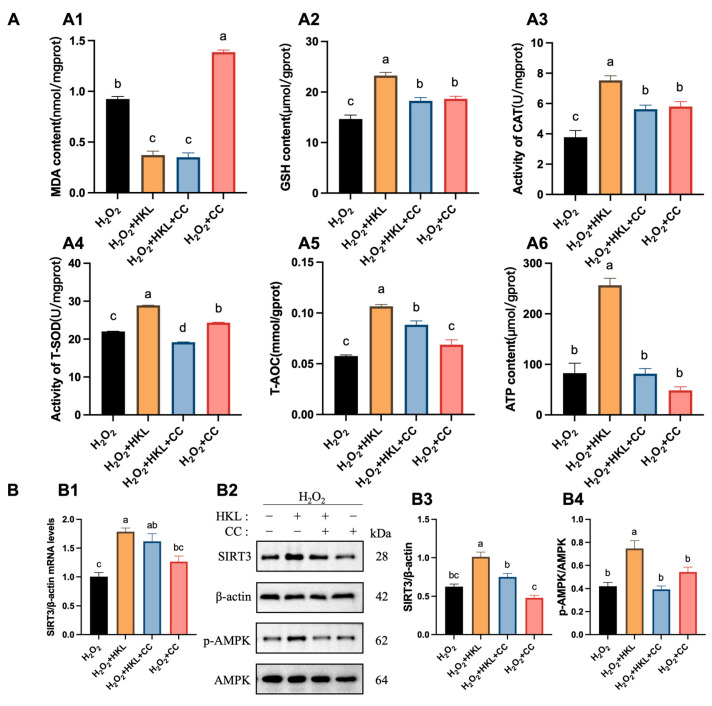
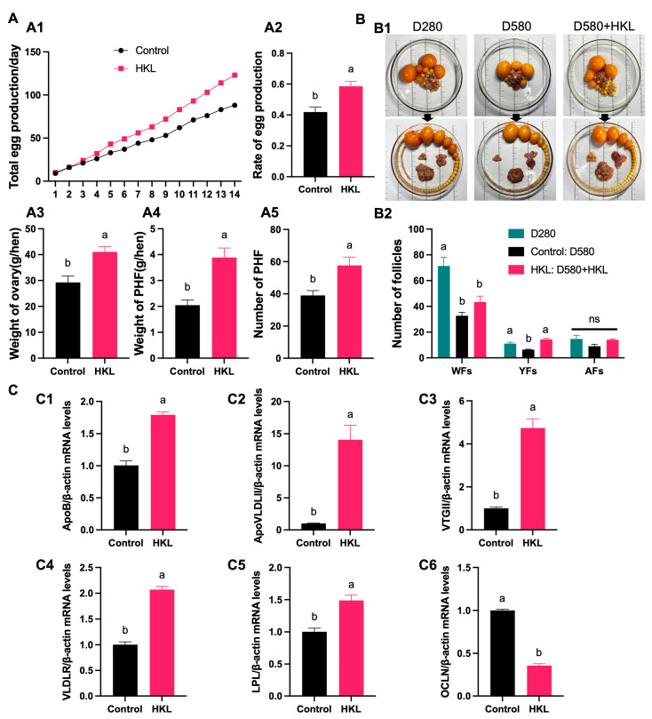
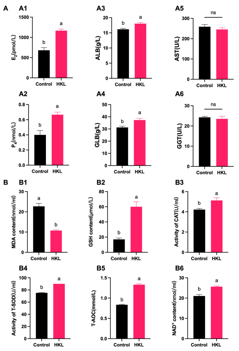
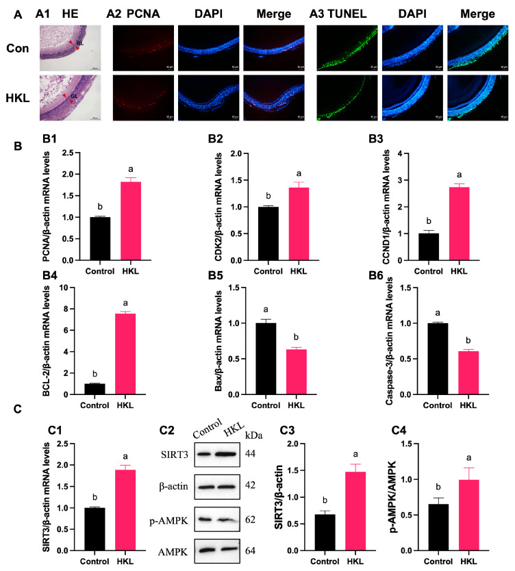
Aging is not only a key internal cause of age-related diseases in humans but also poses a threat to the productivity of farm animals with longer breeding cycles, such as laying chickens. Various measures were taken to prolong the laying period by reducing oxidative stress to improve poultry ovarian functions. Within the mitochondria, SIRT3, a member of the Sirtuin family, plays an important role in post-translational modifications and the regulation of protein activities involved in energy metabolism and oxidative response. This study aimed to investigate the alleviating effect of a bioactive lignan Honokiol (HKL) on oxidative stress in aging chicken ovaries in order to retard decline in egg production. The results showed that HKL treatment restored the abnormal balance between cell proliferation and apoptosis, and it enhanced the antioxidant capacity of the H2O2-induced small white follicles (SWFs) by activating the SIRT3/AMPK pathway. Moreover, HKL significantly increased total egg production, the number of yellow follicles, and the mRNA expression of yolk synthesis and deposition-related genes, serum estrogen, and antioxidant levels. These findings suggest that HKL holds promise in enhancing the egg productivity of aging laying chickens by promoting yolk deposition and reducing ovarian oxidative stress.
Keywords: AMPK; Honokiol; Sirtuins; chicken; ovarian aging; oxidative stress.
Conflict of interest statement
The authors declare no conflicts of interest.
Figures

Similar articles
-
Pterostilbene, a Resveratrol Derivative, Improves Ovary Function by Upregulating Antioxidant Defenses in the Aging Chickens via Increased SIRT1/Nrf2 Expression.Antioxidants (Basel). 2024 Aug 1;13(8):935. doi: 10.3390/antiox13080935. Antioxidants (Basel). 2024. PMID: 39199181 Free PMC article.
-
Flavonoid Fisetin Alleviates Ovarian Aging of Laying Chickens by Enhancing Antioxidant Capacity and Glucose Metabolic Homeostasis.Antioxidants (Basel). 2024 Nov 21;13(12):1432. doi: 10.3390/antiox13121432. Antioxidants (Basel). 2024. PMID: 39765761 Free PMC article.
-
Nobiletin Ameliorates Aging of Chicken Ovarian Prehierarchical Follicles by Suppressing Oxidative Stress and Promoting Autophagy.Cells. 2024 Feb 27;13(5):415. doi: 10.3390/cells13050415. Cells. 2024. PMID: 38474379 Free PMC article.
-
Mitigation of honokiol on fluoride-induced mitochondrial oxidative stress, mitochondrial dysfunction, and cognitive deficits through activating AMPK/PGC-1α/Sirt3.J Hazard Mater. 2022 Sep 5;437:129381. doi: 10.1016/j.jhazmat.2022.129381. Epub 2022 Jun 16. J Hazard Mater. 2022. PMID: 35752048
-
Sirtuins in gamete biology and reproductive physiology: emerging roles and therapeutic potential in female and male infertility.Hum Reprod Update. 2018 May 1;24(3):267-289. doi: 10.1093/humupd/dmy003. Hum Reprod Update. 2018. PMID: 29447380 Review.
Cited by
-
Pterostilbene, a Resveratrol Derivative, Improves Ovary Function by Upregulating Antioxidant Defenses in the Aging Chickens via Increased SIRT1/Nrf2 Expression.Antioxidants (Basel). 2024 Aug 1;13(8):935. doi: 10.3390/antiox13080935. Antioxidants (Basel). 2024. PMID: 39199181 Free PMC article.
-
Dietary Puerariae Lobatae Radix polysaccharides improve ovarian function and reproductive efficiency in laying hens with fatty liver hemorrhagic syndrome.Poult Sci. 2025 May;104(5):105062. doi: 10.1016/j.psj.2025.105062. Epub 2025 Mar 18. Poult Sci. 2025. PMID: 40120252 Free PMC article.
-
Attenuating Effect of a Polyphenol Ellagic Acid on Ovarian Aging by Inhibiting the Ferroptosis Pathway in Low-Yield Laying Chickens.Antioxidants (Basel). 2025 May 21;14(5):614. doi: 10.3390/antiox14050614. Antioxidants (Basel). 2025. PMID: 40427495 Free PMC article.
-
Dietary Saccharomyces cerevisiae fermentation product improved egg quality by modulating intestinal health, ovarian function, and cecal microbiota in post-peak laying hens.Poult Sci. 2025 May;104(5):104979. doi: 10.1016/j.psj.2025.104979. Epub 2025 Mar 7. Poult Sci. 2025. PMID: 40073632 Free PMC article.
-
SIRT3 mediates CPT2 delactylation to enhance mitochondrial function and proliferation in goat granulosa cells.J Anim Sci Biotechnol. 2025 Jul 17;16(1):101. doi: 10.1186/s40104-025-01231-8. J Anim Sci Biotechnol. 2025. PMID: 40671144 Free PMC article.
References
Grants and funding
LinkOut - more resources
Full Text Sources
