. 2024 Mar 21;13(3):383.
doi: 10.3390/antiox13030383.

Cyanide Insensitive Oxidase Confers Hydrogen Sulfide and Nitric Oxide Tolerance to Pseudomonas aeruginosa Aerobic Respiration

Affiliations

Cyanide Insensitive Oxidase Confers Hydrogen Sulfide and Nitric Oxide Tolerance to Pseudomonas aeruginosa Aerobic Respiration

Martina R Nastasi et al. Antioxidants (Basel). .

Abstract

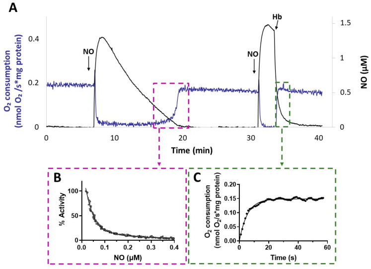
Hydrogen sulfide (H2S) and nitric oxide (NO) are long-known inhibitors of terminal oxidases in the respiratory chain. Yet, they exert pivotal signaling roles in physiological processes, and in several bacterial pathogens have been reported to confer resistance against oxidative stress, host immune responses, and antibiotics. Pseudomonas aeruginosa, an opportunistic pathogen causing life-threatening infections that are difficult to eradicate, has a highly branched respiratory chain including four terminal oxidases of the haem-copper type (aa3, cbb3-1, cbb3-2, and bo3) and one oxidase of the bd-type (cyanide-insensitive oxidase, CIO). As Escherichia coli bd-type oxidases have been shown to be H2S-insensitive and to readily recover their activity from NO inhibition, here we tested the effect of H2S and NO on CIO by performing oxygraphic measurements on membrane preparations from P. aeruginosa PAO1 and isogenic mutants depleted of CIO only or all other terminal oxidases except CIO. We show that O2 consumption by CIO is unaltered even in the presence of high levels of H2S, and that CIO expression is enhanced and supports bacterial growth under such stressful conditions. In addition, we report that CIO is reversibly inhibited by NO, while activity recovery after NO exhaustion is full and fast, suggesting a protective role of CIO under NO stress conditions. As P. aeruginosa is exposed to H2S and NO during infection, the tolerance of CIO towards these stressors agrees with the proposed role of CIO in P. aeruginosa virulence.

Keywords: Pseudomonas aeruginosa; bd-type terminal oxidases; cyanide insensitive oxidase; hydrogen sulfide; nitric oxide; respiratory chain.


Conflict of interest statement

The authors declare no conflicts of interest.

Figures

Figure 1
Figure 1
O2 consumption by membrane preparations from P. aeruginosa mutants sustained with different reducing systems. O2 consumption activity of membranes prepared from the Δcio and ΔcyoΔccoΔcox mutants, measured at 25 °C in the presence of NADH (0.5 mM), ascorbate/TMPD (2 mM/0.2 mM), or DTT/Q1 (10 mM/0.25 mM). The average of at least three different experiments is reported together with the SD. For each condition, significant differences between the Δcio and ΔcyoΔccoΔcox mutants are indicated with asterisks (**, p < 0.01; ****, p < 0.0001).
Figure 2
Figure 2
Effect of sulfide (H2S) on NADH-mediated O2 consumption activity of membranes from P. aeruginosa Δcio and ΔcyoΔccoΔcox. Representative oxygraphic traces collected at 25 °C in the presence of 1 mM NADH before and after addition of 78 µM sulfide to membrane preparations of Δcio (A) or ΔcyoΔccoΔcox (B) mutants. Blue line: O2 concentration. Black line: O2 consumption rate. The protein concentrations of the Δcio and ΔcyoΔccoΔcox membranes were equal to 0.20 and 0.11 mg/mL, respectively.
Figure 3
Figure 3
Effect of sulfide on O2 consumption activity of membranes from P. aeruginosa Δcio and ΔcyoΔccoΔcox. O2 consumption activity of membrane preparations of the Δcio and ΔcyoΔccoΔcox strains measured in the presence of (A) NADH (1 mM), (B) DTT/Q1 (10 mM/0.25 mM), or (C) ascorbate/TMPD (2 mM/0.2 mM) at increasing concentrations of sulfide. The average of three independent experiments is reported with the SD. Asterisks (*, p < 0.05; **, p < 0.01; ***, p < 0.001) denote significant differences between ΔcyoΔccoΔcox and ∆cio (panels (A,B), black asterisks) or with respect to the control not treated with sulfide (panels (AC), red asterisks).
Figure 4
Figure 4
Effect of NaHS on P. aeruginosa cell growth. (A) Growth curves of the indicated strains incubated at 37 °C with shaking (200 rpm) in Lysogeny Broth supplemented with 4 mM L-cysteine (LB-cys) in the absence (solid lines, full circles; untreated samples) or presence of 200 µM NaHS (dashed lines, empty circles; treated samples). The average of three independent experiments is reported with the SD. Differences between treated and untreated cultures of both PAO1 and ∆cio were statistically significant after 2 h incubation (p < 0.001). (B) Fold change in cioA mRNA level in PAO1 grown in LB-cys supplemented with 200 µM NaHS relative to the same strain grown in LB-cys alone. The average of three independent experiments is reported with the SD (*, p < 0.05).
Figure 5
Figure 5
Effect of nitric oxide (NO) on NADH-mediated O2 consumption activity of membranes of P. aeruginosa mutants Δcio and ΔcyoΔccoΔcox. Representative oxygraphic experiments performed at 25 °C in the presence of 1 mM NADH with membrane preparations of the Δcio (A) and ΔcyoΔccoΔcox (B) strains. [NO] = 1.6 µM. The protein concentration of the Δcio and ΔcyoΔccoΔcox membranes was equal to 0.27 and 0.23 mg/mL, respectively.
Figure 6
Figure 6
O2 consumption activity recovered after NO inhibition of respiration of P. aeruginosa Δcio and ΔcyoΔccoΔcox membranes. O2 reductase activity recovered after NO inhibition of respiration of membranes prepared from the indicated mutants measured in the presence of (A) NADH (1 mM), (B) DTT/ Q1 (10 mM/0.25 mM), or (C) ascorbate/TMPD (2 mM/0.2 mM). The average of three independent experiments is reported with the SD. Asterisks (*, p < 0.05; **, p < 0.01) denote significant differences between ΔcyoΔccoΔcox and ∆cio (panels (A,B)) or with respect to the control untreated with NO (panel (C)).
Figure 7
Figure 7
NO inhibition of CIO. (A) O2 consumption rate (blue line) and NO concentration (black line) simultaneously recorded after addition of NO to ΔcyoΔccoΔcox membranes respiring O2 in the presence of NADH. Following the addition of the first aliquot of NO, administered at 100 μM O2, respiration is transiently and fully inhibited. As NO slowly decays by reaction with O2 in solution, respiration recovery takes place, eventually reaching the rate observed prior to inhibition. Afterwards, addition of a second aliquot of NO at 80 μM O2 inhibits respiration again. Following the addition of excess HbO2, which rapidly reacts with NO, fast activity recovery is observed upon NO disappearance from solution. Additions: NADH (1 mM), CIO-containing membranes (0.2 mg protein/mL), NO (1.3 µM), HbO2 (15 μM). (B) Percentage of control activity measured as NO vanishes from solution. Data from the relative boxed area depicted in panel A are plotted as a function of [NO], and data analysis yielded a half-maximal inhibitory concentration for NO value of 55 nM in this sample. The data do not intercept the y-axis, as invariably observed and tentatively explained by limitations in the NO electrode response at very low NO concentrations. (C) Respiration rates measured after addition of HbO2. Data from the relative boxed area depicted in panel (A) are plotted as a function of time and fitted to a single exponential (solid line), yielding an NO off-rate value of 0.18 s−1.

Similar articles

Cited by

References

    1. Szabo C. A Timeline of Hydrogen Sulfide (H2S) Research: From Environmental Toxin to Biological Mediator. Biochem. Pharmacol. 2018;149:5–19. doi: 10.1016/j.bcp.2017.09.010. - DOI - PMC - PubMed
    1. Deshmukh R., Harwansh R.K., Bandyopadhyay N., Bandopadhyay S., Kumar P. Frontiers in Pharmacology of Neurotransmitters. Springer; Singapore: 2020. Pharmacology of Gasotransmitters (Nitric Oxide and Carbon Monoxide) and Their Action; pp. 579–617.
    1. Lo Faro M.L., Fox B., Whatmore J.L., Winyard P.G., Whiteman M. Hydrogen Sulfide and Nitric Oxide Interactions in Inflammation. Nitric Oxide. 2014;41:38–47. doi: 10.1016/j.niox.2014.05.014. - DOI - PubMed
    1. Mancuso C., Navarra P., Preziosi P. Roles of Nitric Oxide, Carbon Monoxide, and Hydrogen Sulfide in the Regulation of the Hypothalamic–Pituitary–Adrenal Axis. J. Neurochem. 2010;113:563–575. doi: 10.1111/j.1471-4159.2010.06606.x. - DOI - PubMed
    1. Farrugia G., Szurszewski J.H. Carbon Monoxide, Hydrogen Sulfide, and Nitric Oxide as Signaling Molecules in the Gastrointestinal Tract. Gastroenterology. 2014;147:303–313. doi: 10.1053/j.gastro.2014.04.041. - DOI - PMC - PubMed