. 2024 Feb 23;12(3):503.
doi: 10.3390/biomedicines12030503.

Targeting Oral Squamous Cell Carcinoma with Combined Polo-Like-Kinase-1 Inhibitors and γ-Radiation Therapy

Affiliations

Targeting Oral Squamous Cell Carcinoma with Combined Polo-Like-Kinase-1 Inhibitors and γ-Radiation Therapy

Subhanwita Sarkar et al. Biomedicines. .

Abstract

Polo-like-kinase-1 (PLK-1) is a serine/threonine kinase that regulates the cell cycle and acts as an oncogene in multiple cancers, including oral squamous cell carcinoma (OSCC). The loss of PLK-1 can inhibit growth and induce apoptosis, making it an attractive therapeutic target in OSCC. We evaluated the efficacy of PLK-1 inhibitors as novel, targeted therapeutics in OSCC. PLK-1 inhibition using BI6727 (volasertib) was found to affect cell death at low nanomolar concentrations in most tested OSCC cell lines, but not in normal oral keratinocytes. In cell lines resistant to volasertib alone, pre-treatment with radiotherapy followed by volasertib reduced cell viability and induced apoptosis. The combinatorial efficacy of volasertib and radiotherapy was replicated in xenograft mouse models. These findings highlight the potential of adding PLK-1 inhibitors to adjuvant therapy regimens in OSCC.

Keywords: OSCC; polo-like-kinase-1; volasertib; γ-radiation therapy.


Conflict of interest statement

The authors declare no conflicts of interest.

Figures
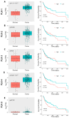
Figure 1
Figure 1
PLK expression in OSCC and correlation to patient survival. (A) PLK-1 is significantly upregulated in OSCC tumor samples when compared to normal oral epithelial tissue, and high PLK-1 expression is correlated to significantly poor overall OSCC patient survival. (B) PLK-2 is significantly upregulated in OSCC tumor samples when compared to normal oral epithelial tissue; however, PLK-2 expression is not correlated to overall OSCC patient survival. (C) PLK-3 expression is not significantly altered in OSCC tumor samples when compared to normal oral epithelial tissue, and high PLK-3 expression is not correlated to overall OSCC patient survival. (D) PLK-4 is significantly upregulated in OSCC tumor samples when compared to normal oral epithelial tissue, and high PLK-4 expression is correlated to significantly poor overall OSCC patient survival. (E) PLK-5 expression is not significantly altered in OSCC tumor samples when compared to normal oral epithelial tissue; however, high PLK-5 expression is correlated to significantly poor overall OSCC patient survival.
Figure 2
Figure 2
Therapeutic efficacy of PLK-1 inhibitors in human cells. (A) The normal oral keratinocyte immortalized with telomerase (OKF6/TERT2) and normal skin human diploid fibroblast (Hs68) cells treated with BI2536 and BI6727 (volasertib) only showed modest cell toxicity. (B) The majority of OSCC cells tested showed significant toxicity with PLK-1 inhibition at nanomolar concentrations. However, UMSCC7 and UMSCC43 were almost completely resistant to PLK-1 inhibition.
Figure 3
Figure 3
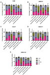
Cell cycle analysis of OSCC cells after volasertib treatment. (A) The majority of the Hs68 fibroblasts progressed through the cell cycle even after treatment with volasertib. (B,C) Volasertib-sensitive CAL27 and UMSCC1 cell lines were arrested at G2/M with treatment. (D,E) Volasertib-resistant UMSCC7 and UMSCC43 cell lines behaved in a similar way to Hs68 cells, escaping volasertib-induced G2/M arrest at different time points.
Figure 4
Figure 4
Volasertib induces apoptosis in sensitive cell lines. (A,B) Lysates from volasertib-sensitive cell lines CAL27 and UMSCC1 showed reduction in p-PLK-1 levels and induction of apoptosis pathways, as evidenced by cleaved PARP and caspase-3 immunoreactive signals. (C,D) Lysates from the volasertib-resistant cell lines UMSCC7 and UMSCC43 showed maintenance of p-PLK-1 levels and the non-induction of apoptosis pathways, evidenced by the lack of cleaved PARP and caspase-3 immunoreactive signals.
Figure 5
Figure 5
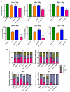
Combination treatment of radiation with adjuvant volasertib. (A) In the volasertib-resistant UMSCC43 cell line, ionising radiation (IR) promotes cytotoxicity. After 48 h of volasertib treatment after 4 Gy of IR, there was a significant reduction in cell viability compared to IR alone. After 72 h of combinatorial treatment, a significant reduction in cell viability was observed at both 2 and 4 Gy of IR. (B) Cell cycle analysis shows a significant increase in cells in the S-phase at 48 h after IR; this effect was further enhanced in the presence of volasertib. (*** p < 0.001).
Figure 6
Figure 6
Effect of combination IR and volasertib treatment in vivo. (A) Adjuvant treatment of volasertib with IR in xenograft mouse tumors led to a significantly smaller tumor size (nadir size) when compared to IR alone. (B) The nadir size was reached slower in the IR and IR + volasertib treatment groups compared to the control or volasertib alone groups. (C) There was no significant change in survival with or without adjuvant volasertib with IR treatment. (D) The tumor volume increase was significantly delayed with IR + volasertib when compared to IR alone.
Figure 7
Figure 7
Schematic representation of the study. (A) A high-throughput drug screen revealed OSCC cells to be sensitive to several PLK-1 inhibitors, including volasertib. (B) OSCC cells can be inherently resistant or sensitive to volasertib. However, exposing the resistant cells with ionization radiation (IR) followed by volasertib leads to significant cell cytotoxicity. (C) In vivo, OSCC cells injected into a flank of NRG mice form tumors that shrink when treated with IR followed by adjuvant volasertib.

Similar articles

References

    1. Tan Y., Wang Z., Xu M., Li B., Huang Z., Qin S., Nice E.C., Tang J., Huang C. Oral squamous cell carcinomas: State of the field and emerging directions. Int. J. Oral Sci. 2023;15:44. doi: 10.1038/s41368-023-00249-w. - DOI - PMC - PubMed
    1. Su Y.-F., Chen Y.-J., Tsai F.-T., Li W.-C., Hsu M.-L., Wang D.-H., Yang C.-C. Current Insights into Oral Cancer Diagnostics. Diagnostics. 2021;11:1287. doi: 10.3390/diagnostics11071287. - DOI - PMC - PubMed
    1. Davaatsend O., Altannamar M., Batbayar B., Jagdagsuren U. Factors influencing the 5-year survival rate of oral cancer patients in the Mongolian population: A retrospective cohort study. Front. Oral Health. 2023;4:1292720. doi: 10.3389/froh.2023.1292720. - DOI - PMC - PubMed
    1. Saka-Herrán C., Jané-Salas E., Mari-Roig A., Estrugo-Devesa A., López-López J. Time-to-Treatment in Oral Cancer: Causes and Implications for Survival. Cancers. 2021;13:1321. doi: 10.3390/cancers13061321. - DOI - PMC - PubMed
    1. Ishigami T., Uzawa K., Higo M., Nomura H., Saito K., Kato Y., Nakashima D., Shiiba M., Bukawa H., Yokoe H., et al. Genes and molecular pathways related to radioresistance of oral squamous cell carcinoma cells. Int. J. Cancer. 2007;120:2262–2270. doi: 10.1002/ijc.22561. - DOI - PubMed

LinkOut - more resources