

Corneal Sub-Basal Nerve Plexus Regeneration Pattern following Implantable Collamer Lens Implantation for Myopia: A Prospective Longitudinal In Vivo Confocal Microscopy Study
- PMID: 38540168
- PMCID: PMC10968349
- DOI: 10.3390/biomedicines12030555
Corneal Sub-Basal Nerve Plexus Regeneration Pattern following Implantable Collamer Lens Implantation for Myopia: A Prospective Longitudinal In Vivo Confocal Microscopy Study
Abstract
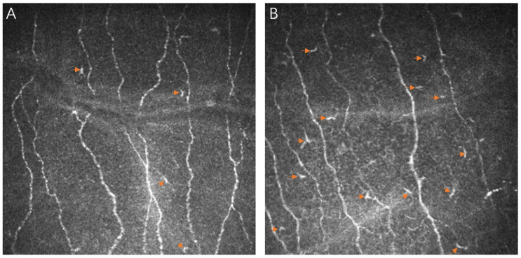
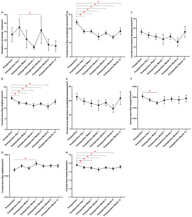
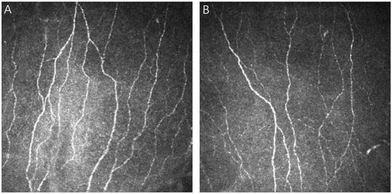
Implantable Collamer Lens (ICL) surgery has increasingly been adopted for myopia correction in recent decades. This study, employing in vivo confocal microscopy (IVCM), aimed to assess the impact of corneal incision during ICL surgery on the corneal sub-basal nerve plexus (SNP) and adjacent immune dendritiform cells (DCs). In this longitudinal study, eyes from 53 patients undergoing ICL surgery were assessed preoperatively and postoperatively over a twelve-month period. Quantification of seven SNP parameters was performed using ACCMetrics V.2 software. Ultimately, the final analysis was restricted to one eye from each of the 37 patients who completed a minimum of three months' postoperative follow-up. Preoperative investigations revealed a positive correlation of DC density with patient age and a negative association with corneal nerve fiber density (CNFD). Additionally, both DCs and CNFD were positively linked to spherical equivalent refraction (SER) and inversely related to axial length (AL). Intriguingly, preoperative DC density demonstrated an indirect relationship with both baseline and postoperative CNFD changes. Post-surgery, an initial surge in DC density was observed, which normalized subsequently. Meanwhile, parameters like CNFD, corneal nerve fiber length (CNFL), and corneal nerve fractal dimension (CNFrD) initially showed a decline following surgery. However, at one-year follow-up, CNFL and CNFrD displayed significant recovery, while CNFD did not return to its baseline level. This study thus delineates the regeneration pattern of SNP and alterations in DC density post-ICL surgery, highlighting that CNFD in the central cornea does not completely revert to preoperative levels within a year. Given these findings, practitioners are advised to exercise caution in older patients, those with high myopia, or elevated preoperative DCs who may undergo delayed SNP regeneration.
Keywords: Visian ICL V4c; corneal confocal microscopy; corneal sub-basal nerve plexus; implantable collamer lens; refractive surgery.
Conflict of interest statement
The authors declare no conflicts of interest.
Figures
Similar articles
-
Comprehensive assessment of corneal microstructural changes following V4c implantable collamer lens surgery using in vivo confocal microscopy.BMC Ophthalmol. 2024 Sep 19;24(1):408. doi: 10.1186/s12886-024-03677-2. BMC Ophthalmol. 2024. PMID: 39300374 Free PMC article.
-
Impact of corneal sub-basal nerve plexus on epithelial thickness after small incision lenticule extraction (SMILE): a quantitative assessment using in vivo confocal microscopy and optical coherence tomography.Quant Imaging Med Surg. 2025 Feb 1;15(2):1254-1264. doi: 10.21037/qims-24-1887. Epub 2025 Jan 22. Quant Imaging Med Surg. 2025. PMID: 39995743 Free PMC article.
-
Longitudinal Morphometric Analysis of Sub-Basal Nerve Plexus in Contralateral Eyes of Patients with Unilateral Neurotrophic Keratitis.Curr Eye Res. 2019 Oct;44(10):1047-1053. doi: 10.1080/02713683.2019.1623899. Epub 2019 Jun 4. Curr Eye Res. 2019. PMID: 31125269
-
In vivo confocal microscopy morphometric analysis of corneal subbasal nerve plexus in dry eye disease using newly developed fully automated system.Graefes Arch Clin Exp Ophthalmol. 2019 Mar;257(3):583-589. doi: 10.1007/s00417-018-04225-7. Epub 2019 Jan 14. Graefes Arch Clin Exp Ophthalmol. 2019. PMID: 30637452
-
Retinal and Choroidal Changes Following Implantable Collamer Lens V4c Implantation in High Myopia Patients-A 1-Year Follow-Up Study.Diagnostics (Basel). 2023 Sep 29;13(19):3097. doi: 10.3390/diagnostics13193097. Diagnostics (Basel). 2023. PMID: 37835840 Free PMC article.
References
-
- Tamimi A., Sheikhzadeh F., Ezabadi S.G., Islampanah M., Parhiz P., Fathabadi A., Poudineh M., Khanjani Z., Pourmontaseri H., Orandi S., et al. Post-LASIK dry eye disease: A comprehensive review of management and current treatment options. Front. Med. 2023;10:1057685. doi: 10.3389/fmed.2023.1057685. - DOI - PMC - PubMed
LinkOut - more resources
Full Text Sources
