. 2024 Mar 6;12(3):586.
doi: 10.3390/biomedicines12030586.

Amelioration of Morphological Pathology in Cardiac, Respiratory, and Skeletal Muscles Following Intraosseous Administration of Human Dystrophin Expressing Chimeric (DEC) Cells in Duchenne Muscular Dystrophy Model

Affiliations

Amelioration of Morphological Pathology in Cardiac, Respiratory, and Skeletal Muscles Following Intraosseous Administration of Human Dystrophin Expressing Chimeric (DEC) Cells in Duchenne Muscular Dystrophy Model

Maria Siemionow et al. Biomedicines. .

Abstract

Duchenne Muscular Dystrophy (DMD) is a lethal disease caused by mutation in the dystrophin gene. Currently there is no cure for DMD. We introduced a novel human Dystrophin Expressing Chimeric (DEC) cell therapy of myoblast origin and confirmed the safety and efficacy of DEC in the mdx mouse models of DMD. In this study, we assessed histological and morphological changes in the cardiac, diaphragm, and gastrocnemius muscles of the mdx/scid mice after the transplantation of human DEC therapy via the systemic-intraosseous route. The efficacy of different DEC doses was evaluated at 90 days (0.5 × 10⁶ and 1 × 10⁶ DEC cells) and 180 days (1 × 10⁶ and 5 × 10⁶ DEC cells) after administration. The evaluation of Hematoxylin & Eosin (H&E)-stained sectional slices of cardiac, diaphragm, and gastrocnemius muscles included assessment of muscle fiber size by minimal Feret's diameter method using ImageJ software. The overall improvement in muscle morphology was observed in DMD-affected target muscles in both studies, as evidenced by a shift in fiber size distribution toward the wild type (WT) phenotype and by an increase in the mean Feret's diameter compared to the vehicle-injected controls. These findings confirm the long-term efficacy of human DEC therapy in the improvement of overall morphological pathology in the muscles affected by DMD and introduce DEC as a novel therapeutic approach for DMD patients.

Keywords: DEC therapy; Duchenne Muscular Dystrophy (DMD); Dystrophin Expressing Chimeric (DEC) cells; cardiac and skeletal muscle morphology; mdx mice; minimal Feret’s fiber diameter; muscle fibrosis; stem cells; systemic-intraosseous administration.


Conflict of interest statement

M.S. is CMO and shareholder of Dystrogen Therapeutics SA, the company that holds a license for DEC Therapy. The author declares a potential financial conflict of interest. M.S. is the inventor on the patent application filed by the University of Illinois at Chicago related to chimeric cell therapy for Duchenne Muscular Dystrophy (WO/2016/201182). K.S. is CEO and shareholder of Dystrogen Therapeutics Corp. A.H. is the adviser to Dystrogen Therapeutics Corp. The authors declare a potential financial conflict of interest. The authors M.S., K.B., K.Z., P.L., S.B., K.S. and A.H. do not have any non-financial conflicts of interest.

Figures

Figure 1
Figure 1
Representative Image of the Minimal Feret’s Fiber Diameter Measurement. The picture represents the H&E-stained transverse section of the Gastrocnemius Muscle (GM) of the 5 × 10⁶ treatment group at 180 days after DEC therapy administration. The yellow double-sided arrow represents the actual minimal Feret’s fiber diameter, which is the shortest distance between two tangents at opposing borders of the muscle fiber. The tangents are drawn for each fiber and diameter is measured in micrometers [μm] after adjusting the scale in the ImageJ software accordingly, as described earlier. Magnification 20×, scale bar 10 μm, Zeiss Meta confocal microscope (Carl Zeiss, Oberkochen, Germany).
Figure 2
Figure 2
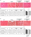
Morphological improvement of the cardiac muscle fibers after 90 and 180 days following the systemic-intraosseous delivery of human DEC therapy. (A) Hematoxylin and Eosin (H&E)-stained histological images representing sectional slices of the mdx/scid mouse heart at 90 days post systemic-intraosseous delivery of DEC cells when compared with the vehicle-injected and wild type (WT) controls. (B) The distribution of myofibers based on the minimal Feret’s diameter measurements revealed a dose-dependent effect of DEC therapy on normalization of the cardiac muscle fiber sizes approaching the WT phenotype. † Significant decrease in the percentage of fibers below 20 μm compared to the group injected with 1 × 10⁶ dose of DEC cells (p < 0.05); ‡ Significant decrease in the percentage of fibers below 20 μm compared to the group injected with 0.5 × 10⁶ dose of DEC cells (p < 0.05) and vehicle-injected controls (p < 0.01); †‡ Significant increase in the percentage of fibers within the 20 μm to 50 μm compared to the group injected with 1 × 10⁶ dose of DEC cells (p < 0.05); ‡‡ Significant increase in the percentage of fibers within 20 μm to 50 μm compared to the group injected with 0.5 × 10⁶ dose of DEC cells (p < 0.05) and vehicle-injected controls (p < 0.01). (C) Mean minimal Feret’s diameter values revealed a significant increase in fiber size in both DEC-injected groups (0.5 × 10⁶ and 1 × 10⁶) when compared to the vehicle-injected controls. (D) Hematoxylin and Eosin (H&E)-stained histological images representing sectional slices of the mdx/scid mouse heart at 180 days post systemic-intraosseous delivery of DEC cells when compared with the vehicle-injected and wild type (WT) controls. (E) The number of fibers based on the minimal Feret’s diameter represents the trend toward the WT phenotype. † Significant decrease in the percentage of fibers below 20 μm compared to the vehicle-injected controls (p < 0.001); ‡ Significant decrease in the percentage of fibers below 20 μm compared to the vehicle-injected controls (p < 0.05); †‡ Significant increase in the percentage of fibers within 20 μm to 50 μm compared to the vehicle-injected controls (p < 0.001); ‡‡ Significant increase in the percentage of fibers within 20 μm to 50 μm compared to the vehicle-injected controls (p < 0.05). (F) Increase in the mean minimal Feret’s diameter of cardiac muscle fibers in mice treated with both DEC therapy doses (1 × 10⁶ cells and 5 × 10⁶ cells) when compared to the vehicle-injected controls. All data presented as means ± SEM. Two-way ANOVA. * p < 0.05, ** p < 0.01. For H&E images on (A,D): magnification 40×, scale bar 50 μm, n = 3/group, and 12 ROI/organ/mouse.
Figure 3
Figure 3
Increase in overall homogeneity of diaphragm after 90 and 180 days following the systemic-intraosseous delivery of human DEC therapy. (A) Hematoxylin and Eosin (H&E)-stained histological images representing sectional slices of the mdx/scid mouse diaphragm at 90 days post systemic-intraosseous delivery of DEC cells when compared with the vehicle-injected and wild type (WT) controls. (B) The distribution of fibers falling into three consecutive increment ranges based on minimal Feret’s diameter showed an increase in the percentage of mid-sized fibers in both DEC-injected groups (0.5 × 10⁶ and 1 × 10⁶) with reduction of very small and large fibers when compared with the vehicle-injected controls. (C) Measurements of the mean minimal Feret’s diameter revealed an increase in mice injected with 0.5 × 10⁶ comparable to the vehicle-injected controls, and an increase approaching the WT phenotype in mice injected with a higher DEC dose (1 × 10⁶). (D) Hematoxylin and Eosin (H&E)-stained histological images representing sectional slices of the mdx/scid mouse diaphragm at 180 days post systemic-intraosseous delivery of DEC cells when compared with the vehicle-injected and wild type (WT) controls. (E) The percentage distribution of myofibers based on the Feret’s diameter showed a decrease in the number of fibers below 20 μm and an increase in the number of larger fibers for the group injected with 1 × 10⁶ dose of DEC cells, with the normalization of diaphragm morphology toward the WT phenotype for mice injected with the dose of 5 × 10⁶ DEC cells, in comparison to the vehicle-injected controls. (F) Mean minimal Feret’s diameter values in mdx/scid mice treated with human DEC therapy revealed no significant increase between both DEC doses and vehicle-injected controls. All data presented as means ± SEM. Two-way ANOVA. For H&E images on (A,D): magnification 20×, scale bar 100 μm, n = 3/ group, and 12 ROI/organ/mouse.
Figure 4
Figure 4
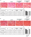
Amelioration of morphological pathology of gastrocnemius muscle after 90 and 180 days following the systemic-intraosseous delivery of human DEC therapy. (A) Hematoxylin and Eosin (H&E)-stained histological images representing sectional slices of the mdx/scid mouse gastrocnemius muscle at 90 days post systemic-intraosseous delivery of DEC cells when compared with the vehicle-injected and wild type (WT) controls. (B) An assessment of fiber size distribution determined by the Feret’s diameter revealed an increase in the percentage of small- and mid-sized fibers in both DEC-injected groups (0.5 × 10⁶ and 1 × 10⁶) with reduction in the number of very large fibers (above 50 μm) when compared with the vehicle-injected controls. (C) Mean minimal Feret’s diameter of gastrocnemius muscle fibers after systemic DEC transplant confirmed normalization of fiber sizes toward WT phenotype in the group injected with 1 × 10⁶ dose of DEC cells. (D) Hematoxylin and Eosin (H&E)-stained histological images representing sectional slices of the mdx/scid mouse gastrocnemius muscle at 180 days post systemic-intraosseous delivery of DEC cells when compared with the vehicle-injected and wild type (WT) controls. (E) The percentage distribution of gastrocnemius muscle fibers depending on the minimal Feret’s diameter revealed a shift toward larger fiber sizes in both treatment groups (1 × 10⁶ and 5 × 10⁶) when compared with the vehicle-injected controls. (F) Measurements of the mean minimal Feret’s diameter showed a significant increase in the mdx/scid mice treated with 5 × 10⁶ dose of DEC therapy when compared with the vehicle-injected controls. All data presented as means ± SEM. Two-way ANOVA. ** p < 0.01. For H&E images on A and D: magnification 20×, scale bar 100 μm, n = 3/group, and 12 ROI/organ/mouse.

References

    1. Van Ruiten H.J., Bettolo C.M., Cheetham T., Eagle M., Lochmuller H., Straub V., Bushby K., Guglieri M. Why are some patients with Duchenne muscular dystrophy dying young: An analysis of causes of death in North East England. Eur. J. Paediatr. Neurol. 2016;20:904–909. doi: 10.1016/j.ejpn.2016.07.020. - DOI - PubMed
    1. Maggio I., Liu J., Janssen J.M., Chen X., Gonçalves M.A.F.V. Adenoviral vectors encoding CRISPR/Cas9 multiplexes rescue dystrophin synthesis in unselected populations of DMD muscle cells. Sci. Rep. 2016;6:37051. doi: 10.1038/srep37051. - DOI - PMC - PubMed
    1. Hoffman E.P., Brown R.H., Kunkel L.M. Dystrophin: The protein product of the duchenne muscular dystrophy locus. Cell. 1987;51:919–928. doi: 10.1016/0092-8674(87)90579-4. - DOI - PubMed
    1. Kharraz Y., Guerra J., Pessina P., Serrano A.L., Muñoz-Cánoves P. Understanding the process of fibrosis in duchenne muscular dystrophy. BioMed Res. Int. 2014;2014:965631. doi: 10.1155/2014/965631. - DOI - PMC - PubMed
    1. Porcari P., Hall M.G., Clark C.A., Greally E., Straub V., Blamire A.M. Time-dependent diffusion MRI as a probe of microstructural changes in a mouse model of Duchenne muscular dystrophy. NMR Biomed. 2020;33:e4276. doi: 10.1002/nbm.4276. - DOI - PubMed

LinkOut - more resources