. 2024 Mar 7;12(3):598.
doi: 10.3390/biomedicines12030598.

An Integrated Approach Including CRISPR/Cas9-Mediated Nanopore Sequencing, Mate Pair Sequencing, and Cytogenomic Methods to Characterize Complex Structural Rearrangements in Acute Myeloid Leukemia

Affiliations

An Integrated Approach Including CRISPR/Cas9-Mediated Nanopore Sequencing, Mate Pair Sequencing, and Cytogenomic Methods to Characterize Complex Structural Rearrangements in Acute Myeloid Leukemia

Michael Phan et al. Biomedicines. .

Abstract

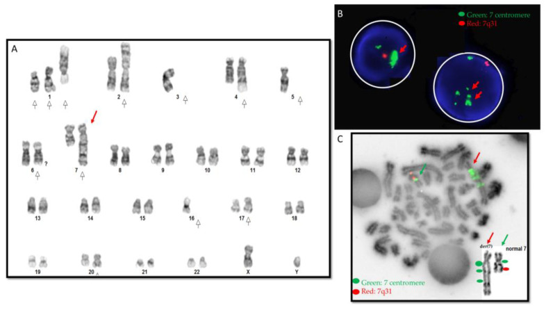
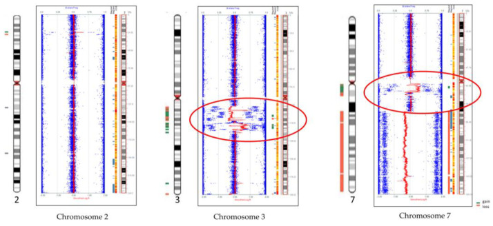
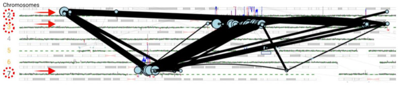
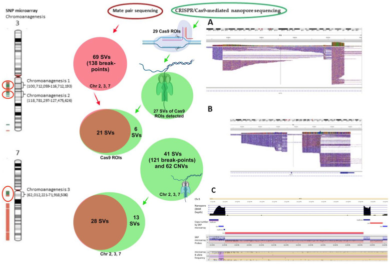
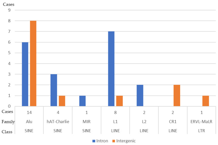
Complex structural chromosome abnormalities such as chromoanagenesis have been reported in acute myeloid leukemia (AML). They are usually not well characterized by conventional genetic methods, and the characterization of chromoanagenesis structural abnormalities from short-read sequencing still presents challenges. Here, we characterized complex structural abnormalities involving chromosomes 2, 3, and 7 in an AML patient using an integrated approach including CRISPR/Cas9-mediated nanopore sequencing, mate pair sequencing (MPseq), and SNP microarray analysis along with cytogenetic methods. SNP microarray analysis revealed chromoanagenesis involving chromosomes 3 and 7, and a pseudotricentric chromosome 7 was revealed by cytogenetic methods. MPseq revealed 138 structural variants (SVs) as putative junctions of complex rearrangements involving chromosomes 2, 3, and 7, which led to 16 novel gene fusions and 33 truncated genes. Thirty CRISPR RNA (crRNA) sequences were designed to map 29 SVs, of which 27 (93.1%) were on-target based on CRISPR/Cas9 crRNA nanopore sequencing. In addition to simple SVs, complex SVs involving over two breakpoints were also revealed. Twenty-one SVs (77.8% of the on-target SVs) were also revealed by MPseq with shared SV breakpoints. Approximately three-quarters of breakpoints were located within genes, especially intronic regions, and one-quarter of breakpoints were intergenic. Alu and LINE repeat elements were frequent among breakpoints. Amplification of the chromosome 7 centromere was also detected by nanopore sequencing. Given the high amplification of the chromosome 7 centromere, extra chromosome 7 centromere sequences (tricentric), and more gains than losses of genomic material, chromoanasynthesis and chromothripsis may be responsible for forming this highly complex structural abnormality. We showed this combination approach's value in characterizing complex structural abnormalities for clinical and research applications. Characterization of these complex structural chromosome abnormalities not only will help understand the molecular mechanisms responsible for the process of chromoanagenesis, but also may identify specific molecular targets and their impact on therapy and overall survival.

Keywords: CRISPR/Cas9; acute myeloid leukemia; chromoanagenesis; complex structural abnormalities; mate pair sequencing; nanopore sequencing.


Conflict of interest statement

The authors declare no conflicts of interest.

Figures

Figure 1
Figure 1
Cytogenetic data. (A) Karyogram. Red arrows point to an abnormal derivative chromosome 7, and white arrows point to other numerical and structural abnormalities. (B) Interphase FISH revealed amplification of chromosome 7 centromeres (in green color, pointed by red arrows) and deletion of 7q31 (in red color). (C) Metaphase FISH. The derivative chromosome 7 (red arrow) shows multiple signals and amplification of the green centromere signal. The green arrow points to a normal chromosome 7. The right-side inserted box shows the make-up of the derivative chromosome 7 by conventional chromosome analysis and FISH data. The derivative chromosome 7 was pseudotricentric and showed centromere amplification.
Figure 2
Figure 2
Genome-wide SNP microarray revealed chromoanagenesis regions on chromosomes 3 and 7 (red circles) and loss of 7q (7q21.11−7q26.3). Chromosome 2 is normal except for two small losses and one gain. Blue dots for genotype (B allele frequency) and red lines for copy number based on probe intensities.
Figure 3
Figure 3
Mate pair sequencing revealed complex rearrangement involving chromosomes 2, 3, and 7. Only structural variants involving these three chromosomes are shown by genome plot (black lines), and breakpoints are shown by solid light green circles. Red arrows pointed to chromosomes 2, 3, and 7.
Figure 4
Figure 4
Summarization of various structural variant breakpoints revealed by CRISPR/Cas9-mediated nanopore sequencing, MPseq, and SNP microarray. From left to right, SNP microarray revealed three chromoanagenesis regions: two on chromosome 3q and one on 7q (shown by red circles). Mate pair sequencing data is in a solid red circle, CRISPR/Cas9 nanopore sequencing data is in green circles, and overlapped data of MPseq and CRISPR/cas9 are in brown circles. (A,B) Complex SVs in the IGV view. Reads of chromosome 7q11.22 genomic region (69,431,166–69,480,285, A) and 3q21.3 genomic region (126,248,059–126,263,338, B). (C) Copy number variants by nanopore sequencing and SNP microarray. Reads of chromosome 3q21.1 genomic region (121,889,250–122,611,342) show complex SVs including amplification, gain, and loss detected by nanopore sequencing, which were consistent with SNP microarray findings. Nanopore reads and SNP microarray data were analyzed by the VIA software to generate copy number variants. Chr: chromosome, CNVs: copy number variants, ROIs: regions of interest by CRISPR/cas9 guideRNAs, SVs: structural variants.
Figure 5
Figure 5
Repeat elements flanking the breakpoints of structural variants in this study. Alu = arthrobactor luteus; CR1 = chicken receptor 1, ERVL = endogenous retrovirus repetitive element, hAT = the hAT superfamily of DNA transposons; LINE = long interspersed nuclear element, L1 = LINE-1, L2 = LINE-2, LTR = long terminal repeat element, MIR = mammalian-wide interspersed repeat, SINE = short interspersed nuclear element.

Similar articles

Cited by

References

    1. Holland A.J., Cleveland D.W. Chromoanagenesis and cancer: Mechanisms and consequences of localized, complex chromosomal rearrangements. Nat. Med. 2012;18:1630–1638. doi: 10.1038/nm.2988. - DOI - PMC - PubMed
    1. Pellestor F. Chromoanagenesis: Cataclysms behind complex chromosomal rearrangements. Mol. Cytogenet. 2019;12:6. doi: 10.1186/s13039-019-0415-7. - DOI - PMC - PubMed
    1. Stephens P.J., Greenman C.D., Fu B., Yang F., Bignell G.R., Mudie L.J., Pleasance E.D., Lau K.W., Beare D., Stebbings L.A., et al. Massive genomic rearrangement acquired in a single catastrophic event during cancer development. Cell. 2011;144:27–40. doi: 10.1016/j.cell.2010.11.055. - DOI - PMC - PubMed
    1. Maher C.A., Wilson R.K. Chromothripsis and human disease: Piecing together the shattering process. Cell. 2012;148:29–32. doi: 10.1016/j.cell.2012.01.006. - DOI - PMC - PubMed
    1. Korbel J.O., Campbell P.J. Criteria for inference of chromothripsis in cancer genomes. Cell. 2013;152:1226–1236. doi: 10.1016/j.cell.2013.02.023. - DOI - PubMed

LinkOut - more resources