Review
. 2024 Mar 14;12(3):649.
doi: 10.3390/biomedicines12030649.

Regenerative Therapy for Corneal Scarring Disorders

Affiliations
Review

Regenerative Therapy for Corneal Scarring Disorders

Christine Chandran et al. Biomedicines. .

Abstract

The cornea is a transparent and vitally multifaceted component of the eye, playing a pivotal role in vision and ocular health. It has primary refractive and protective functions. Typical corneal dysfunctions include opacities and deformities that result from injuries, infections, or other medical conditions. These can significantly impair vision. The conventional challenges in managing corneal ailments include the limited regenerative capacity (except corneal epithelium), immune response after donor tissue transplantation, a risk of long-term graft rejection, and the global shortage of transplantable donor materials. This review delves into the intricate composition of the cornea, the landscape of corneal regeneration, and the multifaceted repercussions of scar-related pathologies. It will elucidate the etiology and types of dysfunctions, assess current treatments and their limitations, and explore the potential of regenerative therapy that has emerged in both in vivo and clinical trials. This review will shed light on existing gaps in corneal disorder management and discuss the feasibility and challenges of advancing regenerative therapies for corneal stromal scarring.

Keywords: cornea scarring; corneal stromal cells; extracellular matrix; gene therapy; regenerative therapy; tissue engineering.


Conflict of interest statement

All authors declare no conflicts of interest. The funders had no role in the study design, data collection and analysis, decision to publish or preparation of the manuscript.

Figures

Figure 1
Figure 1
Human cornea structure and composition. (A) Human cornea anatomy. (B) The human cornea consists of five known layers—three cellular (epithelium, stroma, and endothelium) and two interface non-cellular layers (Bowman’s layer and Descemet’s membrane). (C) The corneal stroma is composed of the extracellular matrix with collagen fibrils organized as stromal lamellae which run orthogonally to each other. Stromal keratocytes are located between stromal lamellae. Transmission electron micrograph with scale bar 2 μm. Created with BioRender.com under license WD26KLB6G7, assessed on 11 March 2024.
Figure 2
Figure 2
A schematic diagram elucidating corneal epithelial–stromal injury and stromal scar formation. In a corneal wound, the damaged epithelium triggers epithelial healing (cell migration and differentiation). The disrupted Bowman’s layer and the infiltration of neutrophils and macrophages allow the invasion of pro-fibrotic growth factors (GFs) and cytokines (CKs) into the stroma. This causes the surviving keratocytes to activate and transit into repair type stromal fibroblasts and contractile myofibroblasts, overproducing ECM proteins with disorganized arrangement and resulting in scar formation. Created with BioRender.com under license JX26IWLDIY, assessed on 1 March 2024.
Figure 3
Figure 3
Causes of corneal haze and opacities.
Figure 4
Figure 4
A schematic diagram of tissue sources of different cell types for corneal stromal regeneration and cell treatment modalities. (A) From ocular corneal tissue, the central stroma derives primary corneal stromal keratocytes (CSKs) for ex vivo culture (Yam 2018 Cell Transplantation). Propagated cells are induced to generate growth-arrested keratocytes expressing different keratocyte-specific gene markers and phenotypes. (B) The anterior limbal stroma derives primary corneal stromal stem cells (CSSCs). The ex vivo expanded cells express various stem cell and MSC gene markers, anti-inflammatory genes, and are capable of generating keratocytes. (C) Extraocular tissues, like adipose, umbilical cord, and bone marrow, are the sources of mesenchymal stem cells (MSCs) with multipotent differentiation potential. (D) Cells are administered to the cornea via topical application in a fibrin gel, intrastromal injection of cell suspension, and intrastromal implantation of cell-ladened tissue or hydrogel scaffolds. Scale bars: 100 μm.
Figure 5
Figure 5
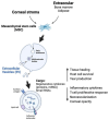
A schematic diagram depicting extracellular vesicles derived from different MSC types and their cargo contents that contribute to corneal tissue regeneration, anti-inflammation, anti-neovascularization, and opacity reduction. Created with BioRender.com under license AU26J1NIGS, assessed on 2 March 2024.
Figure 6
Figure 6
A schematic illustration of bioengineering a biomimetic corneal stroma via integrating native stromal cells (e.g., keratocytes) and ECM-like hydrogel (GelMA). The transplantation to the corneal defect mediates structural restoration of corneal stroma and modulates the stromal matrix environment by proteoglycan secretion to achieve tissue remodeling and scarless wound healing. Created with BioRender.com under license DJ26J1X7F8, assessed on 2 March 2024.

References

    1. Gurtner G.C., Werner S., Barrandon Y., Longaker M.T. Wound repair and regeneration. Nature. 2008;453:314–321. doi: 10.1038/nature07039. - DOI - PubMed
    1. Flaxman S.R., Bourne R.R.A., Resnikoff S., Ackland P., Braithwaite T., Cicinelli M.V., Das A., Jonas J.B., Keeffe J., Kempen J.H., et al. Global causes of blindness and distance vision impairment 1990–2020: A systematic review and meta-analysis. Lancet Glob. Health. 2017;5:e1221–e1234. doi: 10.1016/S2214-109X(17)30393-5. - DOI - PubMed
    1. Jeng B.H., Ahmad S. In Pursuit of the Elimination of Corneal Blindness: Is Establishing Eye Banks and Training Surgeons Enough? Ophthalmology. 2021;128:813–815. doi: 10.1016/j.ophtha.2020.06.042. - DOI - PubMed
    1. Burton M.J. Corneal blindness: Prevention, treatment and rehabilitation. Community Eye Health. 2009;22:33–35. - PMC - PubMed
    1. Sridhar M.S. Anatomy of cornea and ocular surface. Indian J. Ophthalmol. 2018;66:190–194. doi: 10.4103/ijo.IJO_646_17. - DOI - PMC - PubMed

LinkOut - more resources