. 2024 Mar 10;15(3):349.
doi: 10.3390/genes15030349.

A Single-Cell Transcriptome of Bovine Milk Somatic Cells

Affiliations

A Single-Cell Transcriptome of Bovine Milk Somatic Cells

Minja Zorc et al. Genes (Basel). .

Abstract

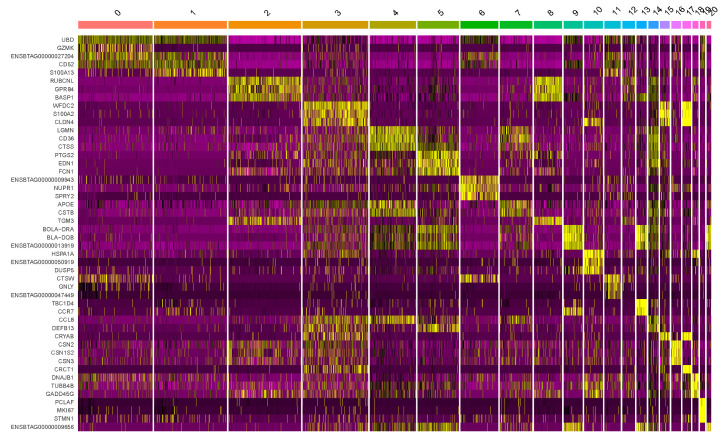
The production of milk by dairy cows far exceeds the nutritional needs of the calf and is vital for the economical use of dairy cattle. High milk yield is a unique production trait that can be effectively enhanced through traditional selection methods. The process of lactation in cows serves as an excellent model for studying the biological aspects of lactation with the aim of exploring the mechanistic base of this complex trait at the cellular level. In this study, we analyzed the milk transcriptome at the single-cell level by conducting scRNA-seq analysis on milk samples from two Holstein Friesian cows at mid-lactation (75 and 93 days) using the 10× Chromium platform. Cells were pelleted and fat was removed from milk by centrifugation. The cell suspension from each cow was loaded on separate channels, resulting in the recovery of 9313 and 14,544 cells. Library samples were loaded onto two lanes of the NovaSeq 6000 (Illumina) instrument. After filtering at the cell and gene levels, a total of 7988 and 13,973 cells remained, respectively. We were able to reconstruct different cell types (milk-producing cells, progenitor cells, macrophages, monocytes, dendritic cells, T cells, B cells, mast cells, and neutrophils) in bovine milk. Our findings provide a valuable resource for identifying regulatory elements associated with various functions of the mammary gland such as lactation, tissue renewal, native immunity, protein and fat synthesis, and hormonal response.

Keywords: cattle; differential gene expression; lactation; mammary gland; milk somatic cells; scRNA-seq; transcriptome.


Conflict of interest statement

The authors declare no conflicts of interest.

Figures

Figure 1
Figure 1
ScRNA−seq quality control analysis with UMAP plots of two somatic milk cell datasets after integration. The colors indicate: (a) number of genes per cell (nFeature_RNA), (b) number of UMI reads per cell (nCount_RNA), (c) percentage of cell counts mapping to mtDNA genes (percent.mt), and (d) percentage of cell counts mapping to ribosomal protein transcripts (percent.rb).
Figure 2
Figure 2
Side-by-side comparison of UMAP projections of cell clusters identified in two samples of fresh bovine milk developed from scRNA-seq. Cells were grouped into 21 matching clusters (labeled with 0 to 20) in Sample 1 and Sample 2.
Figure 3
Figure 3
Annotated cell clusters (n = 21) from two bovine milk samples.
Figure 4
Figure 4
The specificity of marker gene expression was presented with a heatmap of standardized expression for the three major marker genes identified for each of 21 clusters of the cells. The genes are shown in rows and the individual cells in columns. High expression of a particular gene is labeled with yellow and low expression with purple.
Figure 5
Figure 5
Dot plot highlighting the marker genes used to determine the cluster identities of 21 clusters. The marker genes are listed on the X axis and the cluster numbers are on the Y axis. The size of the circle corresponds to the number of cells in the cluster expressing the marker, while the shading corresponds to the extent of expression.
Figure 6
Figure 6
Expression of casein (CSN1S1, CSN1S2, CSN2, and CSN3) and whey protein (PAEP and LALBA) genes in bovine somatic milk cell clusters (labeled with 0 to 20).
Figure 7
Figure 7
Twenty most highly variable expressed genes in bovine milk somatic cells are presented on standardized variance vs. average expression plot. Red dots indicate the differentially expressed genes, and black dots represent the constitutively expressed genes.

References

    1. Inman J.L., Robertson C., Mott J.D., Bissell M.J. Mammary gland development: Cell fate specification, stem cells and the microenvironment. Development. 2015;142:1028–1042. doi: 10.1242/dev.087643. - DOI - PubMed
    1. Neville M.C., McFadden T.B., Forsyth I. Hormonal regulation of mammary differentiation and milk secretion. J. Mammary Gland. Biol. Neoplasia. 2002;7:49–66. doi: 10.1023/A:1015770423167. - DOI - PubMed
    1. Boutinaud M., Jammes H. Potential uses of milk epithelial cells: A review. Reprod. Nutr. Dev. 2002;42:133–147. doi: 10.1051/rnd:2002013. - DOI - PubMed
    1. Wall S.K., Wellnitz O., Bruckmaier R.M., Schwarz D. Differential somatic cell count in milk before, during, and after lipopolysaccharide- and lipoteichoic-acid-induced mastitis in dairy cows. J. Dairy. Sci. 2018;101:5362–5373. doi: 10.3168/jds.2017-14152. - DOI - PubMed
    1. Biteau B., Hochmuth C.E., Jasper H. Maintaining tissue homeostasis: Dynamic control of somatic stem cell activity. Cell Stem Cell. 2011;9:402–411. doi: 10.1016/j.stem.2011.10.004. - DOI - PMC - PubMed

Publication types

LinkOut - more resources