. 2024 Mar 16;15(3):367.
doi: 10.3390/genes15030367.

Dynamics of Mitochondrial DNA Copy Number and Membrane Potential in Mouse Pre-Implantation Embryos: Responses to Diverse Types of Oxidative Stress

Affiliations

Dynamics of Mitochondrial DNA Copy Number and Membrane Potential in Mouse Pre-Implantation Embryos: Responses to Diverse Types of Oxidative Stress

Yasmyn E Winstanley et al. Genes (Basel). .

Abstract

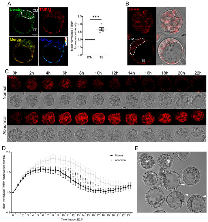
Mitochondria undergo a myriad of changes during pre-implantation embryo development, including shifts in activity levels and mitochondrial DNA (mtDNA) replication. However, how these distinct aspects of mitochondrial function are linked and their responsiveness to diverse stressors is not well understood. Here, we show that mtDNA content increased between 8-cell embryos and the blastocyst stage, with similar copy numbers per cell in the inner cell mass (ICM) and trophectoderm (TE). In contrast, mitochondrial membrane potential (MMP) was higher in TE than ICM. Culture in ambient oxygen (20% O2) altered both aspects of mitochondrial function: the mtDNA copy number was upregulated in ICM, while MMP was diminished in TE. Embryos cultured in 20% O2 also exhibited delayed development kinetics, impaired implantation, and reduced mtDNA levels in E18 fetal liver. A model of oocyte mitochondrial stress using rotenone showed only a modest effect on on-time development and did not alter the mtDNA copy number in ICM; however, following embryo transfer, mtDNA was higher in the fetal heart. Lastly, endogenous mitochondrial dysfunction, induced by maternal age and obesity, altered the blastocyst mtDNA copy number, but not within the ICM. These results demonstrate that mitochondrial activity and mtDNA content exhibit cell-specific changes and are differentially responsive to diverse types of oxidative stress during pre-implantation embryogenesis.

Keywords: inner cell mass; mitochondrial dysfunction; mitochondrial membrane potential; mtDNA copy number.


Conflict of interest statement

The authors declare no conflicts of interest.

Figures

Figure 1
Figure 1
mtDNA copy number during mouse pre-implantation development. (A) Total mtDNA copy number throughout a pre-implantation embryo development time course of IVF-derived embryos (minimum of n = 29 oocytes or embryos analyzed at each developmental stage). (B) Relative mtDNA content per cell in the trophectoderm (TE) and inner cell mass (ICM) micro-dissected from the same blastocysts (n = 48). (C) mtDNA copy number in 2-cell embryos fertilized and cultured in vitro (IVF, n = 141) or fertilized in vivo and collected 40 h following hCG (n = 119). (D) Relative mtDNA content per cell in the ICM from blastocysts fertilized and cultured in vitro (IVF, n = 13) or fertilized in vivo and collected 93 h following hCG (n = 19). (E) Relative mtDNA content per cell in the ICMs of blastocysts on day 5 (n = 13) or day 6 (n = 16) of culture. Data were analyzed using a linear-mixed-effects model (A), where different lowercase letters indicate statistical significance between groups of at least p < 0.05, the paired t-test (B), or the unpaired two-tailed t-test (CE).
Figure 2
Figure 2
Mitochondrial activity from the 8-cell to blastocyst stage differs with embryo developmental competency. (A) In vivo blastocysts from mito-Dendra mice were labeled with TMRM, and the TMRM/Dendra ratio of the ICM and TE was determined (n = 5). (B) In vivo fertilized 8-cell embryos (upper panel) were loaded with TMRM and cultured to the blastocyst stage (lower panel). (C) Representative timelapse images of an 8-cell embryo that exhibited normal development and one that exhibited abnormal development across the 24 h culture period. TMRM (red) is shown in the top panel and brightfield in the bottom. (D) Mean normalized TMRM intensity of embryos that developed into normal (n = 23) versus abnormal (n = 22) blastocysts. (E) Brightfield image of embryos after the 24 h timelapse confocal imaging. Normally developed embryos are indicated by the arrow heads, while abnormal embryos are indicated by the asterisks. Data were analyzed via the paired t-test (A) or two-tailed t-test (D): * p < 0.05, ** p < 0.01, *** p < 0.001, and **** p < 0.0001.
Figure 3
Figure 3
High-oxygen culture delays embryo development and increases ICM mtDNA content. (A) mtROS was detected using the MitoSOX Red Superoxide Indicator in zygotes from control (5%) or high-oxygen (20%) culture. Scale bar is 20 µm. (B) On-time development to the 2-cell stage (n = 12 independent replicates) of embryos cultured in low (5%) or high (20%) oxygen. (C) On-time development to the 8-cell stage (n = 3 independent replicates). (D) Total mtDNA copy number in 8-cell embryos (n = 36 at 5% and n = 47 at 20%). (E) On-time development to the morula stage (n = 4 independent replicates). (F) Total mtDNA copy number in morula embryos (n = 24 at 5% and n = 25 at 20%). (G) On-time development to the blastocyst stage (n = 4 independent replicates). (H) The proportion of blastocysts that were hatching (n = 4 independent replicates). (I) Relative mtDNA per cell in purified ICMs (n = 13 at 5% and n = 12 at 20%). (J) In vivo 8-cell embryos were collected and cultured in the presence of TMRM (10 nM) to the blastocyst stage in either 5% O2 or 20% O2 with TMRM fluorescence in the ICM and TE quantified (n = 8 at 5% and n = 9 at 20%). TE fluorescence normalized to ICM fluorescence. Scale bar is 20µm. (K) Relative mtDNA per cell in fetal (18.5 dpc) liver (n = 30 at 5% and n = 19 at 20%) and (L) fetal heart (n = 30 at 5% and n = 19 at 20%) following blastocyst stage embryo transfer to identical surrogates. Data were analyzed using the paired t-test (B,C,E,G,H) or unpaired t-test (D,F,I,K,L). (J) Data were analyzed via the paired t-test (asterisks) and one-way ANOVA, where different lowercase letters indicate statistical significance of at least p < 0.05: * p < 0.05, ** p = 0.0073, and *** p ≤ 0.0008.
Figure 4
Figure 4
Rotenone-induced mitochondrial dysfunction delays embryo development but does not alter the mtDNA copy number in blastocysts. (A) mtROS was detected using the MitoSOX Red Superoxide Indicator in zygotes from control mice or mice exposed to a low dose of rotenone. (B) Total mtDNA copy number in MII oocytes from control mice or rotenone-exposed females (n = 59 control and n = 60 rotenone). (C) On-time development to the 2-cell stage (n = 23 control and n = 24 rotenone). (D) On-time development to the 8-cell stage (n = 23 control and n = 23 rotenone). (E) Total mtDNA copy number in 8-cell embryos from control or rotenone-exposed females (n = 80 control and n = 61 rotenone). (F) On-time development to the blastocyst stage (n = 23 control and n = 23 rotenone). (G) The proportion of blastocysts that were hatching. (H) Total mtDNA copy number in blastocysts (n = 49 control and n = 72 rotenone). (I) Relative mtDNA per cell in purified ICMs (n = 50 control and n = 43 rotenone). (J) Relative mtDNA per cell in fetal (18.5 dpc) liver (n = 31 control and n = 30 rotenone) and (K) fetal heart (n = 31 control and n = 30 rotenone) following blastocyst stage embryo transfer to identical surrogates. Each data point for on-time development represents the mean of all embryos from one female mouse. Data were analyzed using the unpaired t-test: * p < 0.035, ** p < 0.003, and **** p < 0.0001.
Figure 5
Figure 5
Advanced maternal age and maternal obesity differentially affect blastocyst mtDNA content. (A) mtROS was detected using the MitoSOX Red Superoxide Indicator in MII oocytes from young and reproductive aged (12 months) females. (B) Total mtDNA copy number in MII oocytes from young or aged (n = 110 young and n = 71 aged) female mice. (C) Total mtDNA copy number in 8-cell embryos from young or aged females (n = 39 young and n = 22 aged). (D) Total mtDNA copy number in blastocysts (n = 32 young and n = 13 aged). (E) Relative mtDNA per cell in purified ICMs (n = 82 young and n = 38 aged). (F) mtROS (MitoSOX Red Superoxide Indicator) in zygotes from lean and obese females. (G) total mtDNA copy number in MII oocytes from lean (n = 192 oocytes) and obese (n = 75 oocytes) female mice. (H) Total mtDNA copy number in 8-cell embryos (n = 29 lean, n = 22 obese). (I) Total mtDNA copy number in blastocysts (n = 51 lean, n = 15 obese). (J) Relative mtDNA per cell in purified ICMs (n = 79 lean, n = 61 obese). Data were analyzed using the unpaired t-test: * p < 0.04, ** p = 0.0016, and *** p = 0.0003.

Similar articles

Cited by

References

    1. Dumollard R., Duchen M., Carroll J. The role of mitochondrial function in the oocyte and embryo. Curr. Top. Dev. Biol. 2007;77:21–49. doi: 10.1016/S0070-2153(06)77002-8. - DOI - PubMed
    1. Van Blerkom J. Mitochondrial function in the human oocyte and embryo and their role in developmental competence. Mitochondrion. 2011;11:797–813. doi: 10.1016/j.mito.2010.09.012. - DOI - PubMed
    1. Adhikari D., Lee I.W., Yuen W.S., Carroll J. Oocyte mitochondria-key regulators of oocyte function and potential therapeutic targets for improving fertility. Biol. Reprod. 2022;106:366–377. doi: 10.1093/biolre/ioac024. - DOI - PubMed
    1. Harvey A.J. Mitochondria in early development: Linking the microenvironment, metabolism and the epigenome. Reproduction. 2019;157:R159–R179. doi: 10.1530/REP-18-0431. - DOI - PubMed
    1. Chandel N.S. Mitochondria as signaling organelles. BMC Biol. 2014;12:34. doi: 10.1186/1741-7007-12-34. - DOI - PMC - PubMed

Publication types

LinkOut - more resources