

Roles of Mechanosensitive Channel Piezo1 in Wound Healing and Scar Formation
- PMID: 38541702
- PMCID: PMC10971801
- DOI: 10.3390/life14030377
Roles of Mechanosensitive Channel Piezo1 in Wound Healing and Scar Formation
Abstract
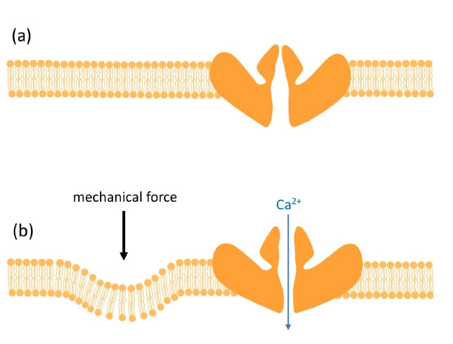
The ability to heal one's wounds is perhaps one of the most fundamental and critical of physiologic processes. This coordinated and closely regulated sequential biological process involves a variety of migratory and resident cells. The activation, modulation, balance, and control of these functions depend upon soluble mediators that activate cells and modulate their diverse functions. Recent advances have identified mechanotransduction as functionally integral in many different cell types and physiologic processes. The mechanically sensitive ion channel Pieoz1 is expressed on platelets, neutrophils, macrophages, endothelial cells, keratinocytes, and fibroblasts, all of which are principally involved in wound healing. On a cellular level, there have been great advances in our understanding of the functional role of Piezo1 mechanotransduction in cutaneous wounding. The blocking of Piezo1 has recently been shown to reduce scarring in vivo and yet, thus far, a comprehensive understanding of the roles that Piezo1 plays in in vivo wound healing remains lacking. Recognizing the ever-present and critical importance of optimal and reparative wound healing, and with the availability of new physical mechanomodulating devices, the time is ripe for gaining deeper insights into optimizing wound healing. In this review, we describe the current knowledge of Piezo1 related to wound healing.
Keywords: Piezo1; ion channel; mechanobiology; scar; wound healing.
Conflict of interest statement
The authors declare no conflicts of interest.
Figures

Similar articles
-
Spatiotemporal dynamics of PIEZO1 localization controls keratinocyte migration during wound healing.Elife. 2021 Sep 27;10:e65415. doi: 10.7554/eLife.65415. Elife. 2021. PMID: 34569935 Free PMC article.
-
Piezo1 in skin wound healing and related diseases: Mechanotransduction and therapeutic implications.Int Immunopharmacol. 2023 Oct;123:110779. doi: 10.1016/j.intimp.2023.110779. Epub 2023 Aug 13. Int Immunopharmacol. 2023. PMID: 37582313 Review.
-
A Piez-o the jigsaw: the Piezo1 channel in skin biology.Clin Exp Dermatol. 2022 Jun;47(6):1036-1047. doi: 10.1111/ced.15138. Epub 2022 Apr 6. Clin Exp Dermatol. 2022. PMID: 35181897 Review.
-
Electricity auto-generating skin patch promotes wound healing process by activation of mechanosensitive ion channels.Biomaterials. 2021 Aug;275:120948. doi: 10.1016/j.biomaterials.2021.120948. Epub 2021 Jun 9. Biomaterials. 2021. PMID: 34157562
-
The Role of the Piezo1 Mechanosensitive Channel in Heart Failure.Curr Issues Mol Biol. 2023 Jul 13;45(7):5830-5848. doi: 10.3390/cimb45070369. Curr Issues Mol Biol. 2023. PMID: 37504285 Free PMC article. Review.
Cited by
-
Immediate Changes in the Elasticity of Tissue and the Pain Pressure Threshold in Cesarean Scar Tissue After a Vacuum Intervention: An Open Clinical Trial.Biomedicines. 2025 Feb 21;13(3):557. doi: 10.3390/biomedicines13030557. Biomedicines. 2025. PMID: 40149534 Free PMC article.
-
Fabrication of Piezo1 protein encapsulated pressure-sensitive multifunctional hydrogel in modulating cellular response and wound healing in pressure ulcer conditions.Regen Ther. 2025 Jul 11;30:371-383. doi: 10.1016/j.reth.2025.06.014. eCollection 2025 Dec. Regen Ther. 2025. PMID: 40689377 Free PMC article.
References
Publication types
LinkOut - more resources
Full Text Sources
