The Value of a Coronary Computed Tomography Angiography plus Stress Cardiac Magnetic Resonance Imaging Strategy for the Evaluation of Patients with Chronic Coronary Syndrome
- PMID: 38541780
- PMCID: PMC10971722
- DOI: 10.3390/jcm13061556
The Value of a Coronary Computed Tomography Angiography plus Stress Cardiac Magnetic Resonance Imaging Strategy for the Evaluation of Patients with Chronic Coronary Syndrome
Abstract
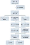
Background: Noninvasive imaging methods, either anatomical or functional tests, serve as essential instruments for the appropriate management of patients with established or suspected coronary artery disease (CAD). We sought to evaluate the safety and efficacy of a coronary computed tomography angiography (CCTA) plus stress cardiac magnetic resonance imaging (S-CMR) strategy in patients with chronic coronary syndrome (CCS). Methods: Patients with suspected CCS showing intermediate coronary plaques (stenosis 30-70%) at CCTA underwent S-CMR. Patients with a positive S-CMR were referred to invasive coronary angiography (ICA) plus instantaneous wave-free ratio (iFR), and myocardial revascularization if recommended. All patients received guideline-directed medical therapy (GDMT), including high-dose statins, regardless of myocardial revascularization. The primary endpoint was a composite of death from cardiovascular causes, non-fatal myocardial infarction, and unplanned revascularization. Results: According to the results of CCTA, 62 patients showing intermediate coronary plaques underwent S-CMR, which was positive for a myocardial perfusion deficit in n = 17 (27%) and negative in n = 45 (73%) patients. According to the results of ICA plus iFR, revascularization was performed in 13 patients. No differences in the primary endpoint between the positive and negative S-CMR groups were observed at 1 year (1 [5.9%] vs. 1 [2.2%], p = 0.485) and after a median of 33.4 months (2 [11.8%] vs. 3 [6.7%]; p = 0.605). Conclusions: Our study suggests that a CCTA plus S-CMR strategy is effective for the evaluation of patients with suspicion of CCS at low-intermediate risk, and it may help to refine the selection of patients with intermediate coronary plaques at CCTA needing coronary revascularization.
Keywords: chronic coronary syndrome; coronary computed tomography angiography; guideline-directed medical therapy; intermediate coronary plaques; revascularization; stress cardiac magnetic resonance imaging.
Conflict of interest statement
The authors declare no conflicts of interest.
Figures


References
-
- Benjamin E.J., Muntner P., Alonso A., Bittencourt M.S., Callaway C.W., Carson A.P., Chamberlain A.M., Chang A.R., Cheng S., Das S.R., et al. Heart Disease and Stroke Statistics-2019 Update: A Report From the American Heart Association. Circulation. 2019;139:e56–e528. doi: 10.1161/CIR.0000000000000659. - DOI - PubMed
-
- Stergiopoulos K., Boden W.E., Hartigan P., Möbius-Winkler S., Hambrecht R., Hueb W., Hardison R.M., Abbott J.D., Brown D.L. Percutaneous coronary intervention outcomes in patients with stable obstructive coronary artery disease and myocardial ischemia: A collaborative meta-analysis of contemporary randomized clinical trials. JAMA Intern. Med. 2014;174:232–240. doi: 10.1001/jamainternmed.2013.12855. - DOI - PubMed
-
- Esposito A., Francone M., Andreini D., Buffa V., Cademartiri F., Carbone I., Clemente A., Guaricci A.I., Guglielmo M., Indolfi C., et al. SIRM-SIC appropriateness criteria for the use of Cardiac Computed Tomography. Part 1: Congenital heart diseases, primary prevention, risk assessment before surgery, suspected CAD in symptomatic patients, plaque and epicardial adipose tissue characterization, and functional assessment of stenosis. Radiol. Med. 2021;126:1236–1248. doi: 10.1007/s11547-021-01378-0. - DOI - PMC - PubMed
LinkOut - more resources
Full Text Sources
Miscellaneous

