Review
. 2024 Mar 15;13(6):1694.
doi: 10.3390/jcm13061694.

Cardiac Sarcoidosis-Diagnostic and Therapeutic Challenges

Affiliations
Review

Cardiac Sarcoidosis-Diagnostic and Therapeutic Challenges

Dennis Korthals et al. J Clin Med. .

Abstract

Sarcoidosis is a multisystem disorder of unknown etiology. The leading hypothesis involves an antigen-triggered dysregulated T-cell-driven immunologic response leading to non-necrotic granulomas. In cardiac sarcoidosis (CS), the inflammatory response can lead to fibrosis, culminating in clinical manifestations such as atrioventricular block and ventricular arrhythmias. Cardiac manifestations frequently present as first and isolated signs or may appear in conjunction with extracardiac manifestations. The incidence of sudden cardiac death (SCD) is high. Diagnosis remains a challenge. For a definite diagnosis, endomyocardial biopsy (EMB) is suggested. In clinical practice, compatible findings in advanced imaging using cardiovascular magnetic resonance (CMR) and/or positron emission tomography (PET) in combination with extracardiac histological proof is considered sufficient. Management revolves around the control of myocardial inflammation by employing immunosuppression. However, data regarding efficacy are merely based on observational evidence. Prevention of SCD is of particular importance and several guidelines provide recommendations regarding device therapy. In patients with manifest CS, outcome data indicate a 5-year survival of around 90% and a 10-year survival in the range of 80%. Data for patients with silent CS are conflicting; some studies suggest an overall benign course of disease while others reported contrasting observations. Future research challenges involve better understanding of the immunologic pathogenesis of the disease for a targeted therapy, improving imaging to aid early diagnosis, assessing the need for screening of asymptomatic patients and randomized trials.

Keywords: cardiac sarcoidosis; cardiovascular magnetic resonance; implantable cardioverter–defibrillator; inflammatory cardiomyopathy; positron emission tomography; sudden cardiac death; ventricular arrhythmia.


Conflict of interest statement

The authors declare no conflicts of interest.

Figures

Figure 1
Figure 1
Two monomorphic VT (A,B) in a 58-year-old female patient with cardiac sarcoidosis and recurrent VT in the presence of significantly reduced RV and LV function, chronic amiodarone therapy and previous implantation of a cardiac resynchronization therapy/defibrillator (CRT-D) system who underwent extensive RV/LV mapping. (C) Successful termination of VT (A) in the anterior right ventricular outflow tract. ABL (ablation catheter). (D) Endocardial voltage map of the RV with extensive low-voltage/scarring (in color 0.5–1.5 mV).
Figure 2
Figure 2
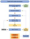
Initial evaluation and selection of patients for advanced imaging, modified from [11]. LBBB: left bundle branch block; RBBB: right bundle branch block; AV: atrioventricular, LVEF: left ventricular ejection fraction; VA: ventricular arrhythmia; CMR: cardiovascular magnetic resonance, FDG-PET: 18-fluordesoxyglucose positron emission tomography; EMB: endomyocardial biopsy.
Figure 3
Figure 3
LGE phenotypes in cardiac sarcoidosis; illustration of a short axis view depicting late gadolinium phenotypes of CS. LGE distribution is most commonly multifocal and patchy. It can affect all myocardial layers (subendocardial, midmyocardial, epicardial) and can also present transmurally. LGE is most frequently seen in the basal septum on the RV side as well as anteroseptal but can appear anywhere. A “hook” sign of septal LGE extending to the RV has been described as marker of CS but is also seen in giant cell myocarditis. The presence of RV LGE might be associated with additional risk for VAs compared to LV involvement only.
Figure 4
Figure 4
LGE-CMR and FDG-PET images for respective short-axis, 4-chamber, and 2-chamber orientations. The patient was a 65-year-old woman with no prior heart condition initially presenting with third-degree AV block. After a year, the patient developed VT; the pacemaker was subsequently upgraded to an ICD system. The diagnosis of CS was established at this time. Top row shows LGE-CMR fibrosis imaging. Arrows indicate areas of abnormal LGE in a patchy distribution pattern reflecting inflammation and/or scar. Bottom row shows fusion FDG-PET/CT showing patchy uptake in regions of scar, suggesting active inflammation. Note the artifact in the RV cavum caused by the ICD lead. LGE: late gadolinium enhancement. FDG: 18-fluordesoxyglucose positron emission tomography.
Figure 5
Figure 5
Proposed treatment algorithm for patients with manifest CS, modified from [10]. * Agents: Methotrexate, Azathioprine, Mycophenolate mofetil; ** we terminate therapy guided by advanced imaging findings; *** agents: Leflunomide, TNF-α-antagonists (Infliximab, Adalimumab), Cyclophosphamide, Rituximab.
Figure 6
Figure 6
(A) Twelve lead ECGs of a 35-year-old male with proven sarcoidosis. Please note the first-degree AV block and the right bundle branch block with a prominent surface area of the maximum R’ wave in leads V1 (see also Hoogendoorn et al. [34]). Monomorphic sustained VT (CL = 400 ms) induced before (B) and 6 months after steroid therapy (C). Before (B) PET demonstrated acute inflammation. This indicates that reduction of inflammation by immunosuppressant therapy may be proarrhythmic. The patient was initially asymptomatic and refused an ICD in (B) which was finally implanted after induction of the faster VT (CL = 270 ms) (C).
Figure 7
Figure 7
Recommendations for prevention of SCD and management of VA in cardiac sarcoidosis, modified from [4]; SCD: sudden cardiac death, VT: ventricular tachycardia, LVEF: left ventricular ejection fraction, ICD: implantable cardioverter–defibrillator, LGE: late gadolinium enhancement; PES: programmed electrical stimulation; SMVT: sustained monomorphic ventricular tachycardia.

References

    1. Drent M., Crouser E.D., Grunewald J. Challenges of Sarcoidosis and Its Management. N. Engl. J. Med. 2021;385:1018–1032. doi: 10.1056/NEJMra2101555. - DOI - PubMed
    1. Kandolin R., Lehtonen J., Airaksinen J., Vihinen T., Miettinen H., Ylitalo K., Kaikkonen K., Tuohinen S., Haataja P., Kerola T., et al. Cardiac sarcoidosis: Epidemiology, characteristics, and outcome over 25 years in a nationwide study. Circulation. 2015;131:624–632. doi: 10.1161/CIRCULATIONAHA.114.011522. - DOI - PubMed
    1. Takaya Y., Nakamura K., Nishii N., Ito H. Clinical outcomes of patients with isolated cardiac sarcoidosis confirmed by clinical diagnostic criteria. Int. J. Cardiol. 2021;345:49–53. doi: 10.1016/j.ijcard.2021.10.150. - DOI - PubMed
    1. Zeppenfeld K., Tfelt-Hansen J., de Riva M., Winkel B.G., Behr E.R., Blom N.A., Charron P., Corrado D., Dagres N., de Chillou C., et al. 2022 ESC Guidelines for the management of patients with ventricular arrhythmias and the prevention of sudden cardiac death. Eur. Heart J. 2022;43:3997–4126. doi: 10.1093/eurheartj/ehac262. - DOI - PubMed
    1. Könemann H., Dagres N., Merino J.L., Sticherling C., Zeppenfeld K., Tfelt-Hansen J., Eckardt L. Spotlight on the 2022 ESC guideline management of ventricular arrhythmias and prevention of sudden cardiac death: 10 novel key aspects. Europace. 2023;25:euad091. doi: 10.1093/europace/euad091. - DOI - PMC - PubMed

LinkOut - more resources