Review
. 2024 Mar 8;25(6):3131.
doi: 10.3390/ijms25063131.

Membrane Interaction Characteristics of the RTX Toxins and the Cholesterol-Dependence of Their Cytolytic/Cytotoxic Activity

Affiliations
Review

Membrane Interaction Characteristics of the RTX Toxins and the Cholesterol-Dependence of Their Cytolytic/Cytotoxic Activity

Helena Ostolaza et al. Int J Mol Sci. .

Abstract

RTX toxins are important virulence factors produced by a wide range of Gram-negative bacteria. They are secreted as water-soluble proteins that are able to bind to the host cell membrane and insert hydrophobic segments into the lipid bilayer that ultimately contribute to the formation of transmembrane pores. Ion diffusion through these pores leads then to cytotoxic and cytolytic effects on the hosts. Several reports have evidenced that the binding of several RTX toxins to the target cell membrane may take place through a high-affinity interaction with integrins of the β2 family that is highly expressed in immune cells of the myeloid lineage. However, at higher toxin doses, cytotoxicity by most RTX toxins has been observed also on β2-deficient cells in which toxin binding to the cell membrane has been proposed to occur through interaction with glycans of glycosylated lipids or proteins present in the membrane. More recently, cumulative pieces of evidence show that membrane cholesterol is essential for the mechanism of action of several RTX toxins. Here, we summarize the most important aspects of the RTX toxin interaction with the target cell membrane, including the cholesterol dependence, the recent identification in the sequences of several RTX toxins of linear motifs coined as the Cholesterol Recognition/interaction Amino acid Consensus (CRAC), and the reverse or mirror CARC motif, which is involved in the toxin-cholesterol interaction.

Keywords: RTX toxins; cholesterol; cholesterol-binding motifs; lipid-protein interactions; pore-forming toxins.


Conflict of interest statement

The authors declare no conflicts of interest.

Figures

Figure 1
Figure 1
General scheme of the synthesis, post-translational modification, and secretion for the RTX toxins. The schematic organization of the operon is represented by boxes labelled from A to D. Gene product A (red) is the polypeptide corresponding to a protoxin (pro-RTX) that matures in the bacterial cytosol to the active form by post-translational acylation at two conserved internal lysine residues [5]. Fatty acylation is mediated by a specific acyltransferase encoded by the product of the gene C (dark green) and an acyl carrier protein (Acyl-ACP) [5,6,7,8,9,10]. The mature, acylated RTX toxin is then directly secreted across both membranes by the type I secretion system (T1SS) constituted by the gene products B (light green) and D (pink) and the bacterial outer membrane TolC protein [11,12,13,14,15]. Adapted from Stanley and cols. [7]. Created with Biorender.com (accessed on 10 January 2024).
Figure 2
Figure 2
Schematic representation of the structure of the RTX toxins UPEC HlyA (top) and B. pertussis CyaA (bottom). These toxins consist of a pore-forming domain (H, dark blue), an acylated segment with two post-translationally acylated lysine residues (indicated with two turquois arrows), a repeat domain (RD dark green), a receptor binding domain (RBD, light blue), and a C-terminal secretion signal (SS, purple). Unlike other RTX toxins, CyaA contains a unique adenylate cyclase AC domain (AC, green) and a translocation region (TR, orange).
Figure 3
Figure 3
Structure of the αMβ2 integrin ectodomain. Structure at 2.7 Å resolution of the heterodimeric αMβ2 integrin published by Goldsmith and cols [62], showing the αM subunit in yellow and the β2 subunit in red. A cartoon representation of the cell membrane lipid bilayer with phospholipid molecules (in purple) and cholesterol molecules (in orange) has been included as well. Figure redrawn from the integrin structure solved by Goldsmith and cols and deposited in PDB ID: 7USL [62].
Figure 4
Figure 4
Schematic representation of the chemical structure of GM1 and GM3 gangliosides.
Figure 5
Figure 5
Chemical structure of cholesterol. Cholesterol has a global conical shape, and it can be divided into two parts. On the one hand, the 4-ring sterane system with the linked hydroxyl group occupies 50% of the spatial volume of the molecule, and it has very low flexibility; on the other, the terminal isooctyl chain is very flexible and, hence, it can adopt numerous conformations when bound to membrane proteins. The combination of polar (3β-hydroxyl group) and apolar (the sterol ring and the isooctyl side chain) regions impart an amphipathic nature to cholesterol, making it conducive to interaction with other membrane components (lipids and proteins). An interesting structural feature of cholesterol is the inherent asymmetry of the sterol ring plane owing to methyl substitutions on one of its faces. The smooth α face is constituted of only axial hydrogen atoms and contributes to favorable van der Waals interaction with the saturated fatty acyl chains of phospholipids. On the other hand, the rough β face characterized by the protruding methyl groups (C18 and C19) can snugly interact with the bumpy topology of a membrane protein.
Figure 6
Figure 6
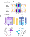
Cholesterol recognition/interaction amino acid consensus motifs. (A) Mirror topology of CRAC/CARC motifs within the same TM domain of a multispanning membrane-protein. The dashed line indicates the border between the inner and outer leaflets. Many cholesterol-binding proteins possess an amino acid sequence in the juxtamembrane region conforming to the pattern (L/V)-X1–5-(Y)-X1–5-(K/R) (CRAC) or the opposite (K/R)-X1–5-(Y/F/W)-X1–5-(L/V) (CARC). (B) Schematic illustration of the interaction between the cholesterol molecule and particular amino acid residues corresponding to CRAC/CARC-like motifs. The CRAC/cholesterol complex displays a parallel head-to-head/tail-to-tail geometry [127,128]. The branched N-terminal Leu or Val apolar residues bind to the isooctyl chain through van der Waals interactions (London forces); the mandatory aromatic residue (Tyr, Phe, or Trp) stacks onto one of the steranes four rings (CH-π stacking); and the C-terminal Lys or Arg polar residues establish hydrogen bonds with the hydroxyl group of CHOL [127]. The aromatic residues are able to stack onto the smooth α face of cholesterol or intercalate between the aliphatic spikes that emerge from the rough β face. The position of the aromatic residue is determined by the length of the X1–5 linkers [127,128]. Redrawn from Fantini and colleagues [127].

Similar articles

References

    1. Welch R.A. Pore-Forming Cytolysins of Gram-Negative Bacteria. Mol. Microbiol. 1991;5:521–528. doi: 10.1111/j.1365-2958.1991.tb00723.x. - DOI - PubMed
    1. Linhartová I., Bumba L., Mašín J., Basler M., Osička R., Kamanová J., Procházková K., Adkins I., Hejnová-Holubová J., Sadílková L., et al. RTX Proteins: A Highly Diverse Family Secreted by a Common Mechanism. FEMS Microbiol. Rev. 2010;34:1076–1112. doi: 10.1111/j.1574-6976.2010.00231.x. - DOI - PMC - PubMed
    1. Welch R.A. RTX Toxin Structure and Function: A Story of Numerous Anomalies and Few Analogies in Toxin Biology. Curr. Top. Microbiol. Immunol. 2001;257:85–111. doi: 10.1007/978-3-642-56508-3_5. - DOI - PubMed
    1. Coote J.G. Structural and Functional Relationships among the RTX Toxin Determinants of Gram-Negative Bacteria. FEMS Microbiol. Rev. 1992;8:137–161. doi: 10.1111/j.1574-6968.1992.tb04961.x. - DOI - PubMed
    1. Stanley P., Packman L.C., Koronakis V., Hughes C. Fatty Acylation of Two Internal Lysine Residues Required for the Toxic Activity of Escherichia coli Hemolysin. Science. 1994;266:1992–1996. doi: 10.1126/science.7801126. - DOI - PubMed

MeSH terms

LinkOut - more resources