. 2024 Mar 9;25(6):3149.
doi: 10.3390/ijms25063149.

Downregulation of Serotonergic System Components in an Experimentally Induced Cryptorchidism in Rabbits

Affiliations

Downregulation of Serotonergic System Components in an Experimentally Induced Cryptorchidism in Rabbits

Francisco Jiménez-Trejo et al. Int J Mol Sci. .

Abstract

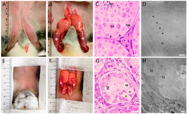
Cryptorchidism (CO) or undescended testes is defined as the failure of one or both testes to be positioned inside the scrotum. Typically, cryptorchidism is detected at birth or shortly thereafter, and in humans, it is considered to be part of the testicular dysgenesis syndrome (TDS), a complex pathology regarding the male reproductive system that apparently involves the interaction of both genetic and environmental harmful factors, mainly during embryonic development. Serotonin (5-HT) is an ancient molecule that participates in a broad range of body functions, and in recent years, its importance in reproduction has started to be elucidated. In male pathologies such as infertility, varicocele, erectile dysfunction, and primary carcinoid tumors, an increase in 5-HT concentration or its metabolites in the blood, semen, and urine has been directly related; nevertheless, the role of 5-HT in CO remains unknown. In the present work, our goal was to answer two important questions: (1) whether some serotonergic system components are present in adult male Oryctolagus cuniculus (chinchilla rabbit) and (2) if there are changes in their expression in an experimental model of CO. Using histological, molecular, and biochemical approaches, we found the presence of some serotonergic system components in the adult chinchilla rabbit, and we demonstrated that its expression is downregulated after CO was pharmacologically induced. Although we did not test the role of 5-HT in the etiology of CO, our results suggest that this indoleamine could be important for the regulation of steroidogenesis and spermatogenesis processes in the chinchilla rabbit during adulthood. Finally, in parallel experimental series, we found downregulation of kynurenine concentration in COI rabbits when compared to control ones, suggesting that CO could be affecting the kynurenine pathway and probably testicular immune privilege which in turn could lead to infertility/sterility conditions in this disorder.

Keywords: cryptorchidism; gonocytes; kynurenine; serotonin; testes.


Conflict of interest statement

The authors declare that no conflicts of interest exist regarding this article.

Figures

Figure 1
Figure 1
Anatomic description of control and COI rabbits. Representative images of intact testes and dissected in control ((A,B) respectively) and COI (E,F) rabbits; asterisks in (B) and (F) denote the testis; absence of both testicular descent and a pigmented scrotal sac is noted in COI rabbits when compared to control ones. (C,G) show representative photomicrographs of testis sections stained with H&E, in control (C) and COI testis (G). Representative TEM microphotograph of 150-day-old normal (D) and COI (H). Please see details in text. (H) S = Sertoli cell; L = Leydig cell; M = peritubular myoid cell; Spg = spermatogonium; Sc1 = primary spermatocyte; Spz = sperm; Ap: atypical cells. G = gonocyte. Scale bar in (C,G): 10 µm, (D): 5 µm, and (H): 7 µm.
Figure 2
Figure 2
Gene expression of some serotonergic system elements in control and COI testes. Relative mRNA levels of Tph1, Maoa, Htr2a, Htr3a, and Slc6a4 (5HTT), in both control and COI rabbits, are shown. * denotes statistical significance differences between experimental conditions. p value was set at ≤0.05.
Figure 3
Figure 3
Immunofluorescence of serotonergic system elements in 150-day-old control rabbit testes’ sections (I). 5-HT was found strongly immunostained in interstitial zone (asterisks), and a weaker signal for it was found in the seminiferous epithelium (A); inset is a panoramic view. TPH1 was found strongly immunostained in clusters of Leydig cells in the interstitium (asterisks); inset shows brain stem neurons used as positive control of technique (B). 5-HT1B receptor (C) was found immunostained in Leydig cells (star), Sertoli-like cells (arrowheads), and the perinuclear region of spermatogonials (arrow). 5-HT2A receptor (D) was found immunostained in spermatogonials (arrowheads), Sertoli-like cells (arrow), secondary spermatocytes (asterisks), and some sperm cytoplasmic droplets in the lumen and was slightly detected in the Leydig cell in interstitium (stars). 5-HT3A receptor (E) was found strongly immunostained in the interstitial zone (star) and in the sperm acrosome (arrowheads); inset shows a panoramic view. MAOA enzyme (F) immunoreactivity was found in Leydig cells (star), spermatogonials (arrows), and putative spermatocytes in preleptotene (arrowheads); inset shows a negative control. Scale bar in (AF): 20 µm.
Figure 4
Figure 4
Immunofluorescence of serotonergic system elements in 150-day-old control rabbit testes’ sections (II). Panoramic view of 5-HTT immunolocalization in the seminiferous tubules, presumptively in Sertoli cells, spermatogonia, spermatocytes, spermatids, and sperm, and in the interstitial zone in the walls of blood vessels ((A); star); higher magnification of a seminiferous tubule (B), in which spermatogonial stem cells show diffuse cytoplasmic staining (arrowheads), Sertoli cells show more vesiculated, diffuse staining (arrow), and both spermatocytes and spermatids show strong cytoplasmic expression (open arrows), presumably in the acrosomal region adjacent to the nucleus; in the same way, spermatozoa (higher magnification at (C)) show strong immunoreactivity in the acrosome (open arrows), and inset shows a negative control; VMAT1 (D) was found strongly stained in the interstitial zone (star) of seminiferous tubules, probably in Sertoli cells (arrowhead), and inset shows a panoramic view at lower magnification. L: lumen of seminiferous tubules. Scale bar in (A): 100 µm and (BD): 10 µm.
Figure 5
Figure 5
Immunofluorescence of serotonergic system elements in 150-day-old COI rabbit testes’ sections (I). Panoramic view of 5-HT1B receptor immunolocalization (A), which was found immunostained all through the small seminiferous tubules in the cell membranes of apparently all cell types belonging to them (arrows) and in clusters of Leydig cells (arrowheads); inset shows a neuron from brain stem used as positive control. 5-HT2A receptor immunoreactivity (B) was found in Sertoli-like cells (arrowheads) and gonocytes (arrow); inset shows a panoramic view at lower magnification. 5-HT3A receptor (C) was found expressed in presumptive gonocytes and Sertoli cells (arrowheads); inset shows a panoramic view at lower magnification. MAOA enzyme (D) was found expressed in all cell layers of the seminiferous tubules (star); inset shows negative control. Scale bar in (A): 100 µm and (BD): 20 µm.
Figure 6
Figure 6
Immunofluorescence of serotonergic system elements in 150-day-old COI rabbit testes’ sections (II). Panoramic view of TPH1 enzyme immunostaining (A), which was found intensely depicted in clusters of Leydig cells (arrowheads) found in the base of testicular tubules (stars); positive neurons from rabbit brain stem are shown in (B), as a positive control, while inset shows negative control; panoramic view of 5-HTT immunostaining (C), in which it was found intensely stained in clusters of Leydig cells (arrows) and cell membranes of gonocytes (arrowheads) and spermatocytes in the seminiferous tubules; higher magnification of a seminiferous tubule in which cell membranes of gonocytes (arrows) and spermatocytes (arrowheads) are intensely stained to 5-HTT is shown in (D). Scale bar in (A,C): 100 µm and (B,D): 20 µm.
Figure 7
Figure 7
Immunofluorescence of serotonergic system elements in 150-day-old COI rabbit testes’ sections (III). Panoramic view of double immunofluorescence for 5-HT and VMAT1 transporter (AC); 5-HT immunoreactivity (A) was mainly distributed along the interstitial zone (stars) and a faintly staining was found in the lumen (L) of testicular tubules,, whereas VMAT1 (B) was distributed in both interstitial zone and seminiferous tubules, apparently in Leydig (stars) and Sertoli cells for the evident cytoplasmic extensions, respectively; apparently, there were some Leydig cells double positive for 5-HT/VMAT1 and another only positive for 5-HT or VMAT1 as shown in the merged image (C); brain stem neurons (arrows) are shown as a positive control for 5-HT in (D). Scale bar in (AD): 100 µm.
Figure 8
Figure 8
Western blotting of some serotonergic system components in control and COI rabbit testes’ homogenates. (A) Representative images for TPH1, MAOA, 5-HT1B, 5-HT2A, 5-HT3A, and VMAT1 immunoblots in control and COI rabbit testes; GAPDH was used as control; (B) densitometric analysis of immunoblots shown on the left. (* p < 0.05, Kruskal–Wallis one-way ANOVA followed by Mann–Whitney U test.)
Figure 9
Figure 9
Comparative model of serotonergic system components in control and COI rabbit testes. The more relevant aspects are the finding of the presence of serotonergic system elements in control rabbits and the downregulation of them in COI rabbits.

References

    1. Lee P.A., Houk C.P., Ahmed S.F., Hughes I.A. Consensus statement on management of intersex disorders. Pediatrics. 2006;118:e488–e500. doi: 10.1542/peds.2006-0738. - DOI - PubMed
    1. Skakkebæk N.E., Rajpert-De Meyts E., Main K.M. Testicular dysgenesis syndrome: An increasingly common developmental disorder with environmental aspects. Hum. Reprod. 2001;16:972–978. doi: 10.1093/humrep/16.5.972. - DOI - PubMed
    1. Serrano T., Chevrier C., Multigner L., Cordier S., Jégou B. International geographic correlation study of the prevalence of disorders of male reproductive health. Hum. Reprod. 2013;28:1974–1986. doi: 10.1093/humrep/det111. - DOI - PubMed
    1. Gore A.C., Holley A.M., Crews D. Mate choice, sexual selection, and endocrine-disrupting chemicals. Horm. Behav. 2018;101:3–12. doi: 10.1016/j.yhbeh.2017.09.001. - DOI - PMC - PubMed
    1. Gurney J.K., McGlynn K.A., Stanley J., Merriman T., Signal V., Shaw C., Edwards R., Richiardi L., Hutson J., Sarfati D. Risk factors for cryptorchidism. Nat. Rev. Urol. 2018;14:534–548. doi: 10.1038/nrurol.2017.90. - DOI - PMC - PubMed