. 2024 Mar 13;25(6):3247.
doi: 10.3390/ijms25063247.

The Role of Neutral Sphingomyelinase-2 (NSM2) in the Control of Neutral Lipid Storage in T Cells

Affiliations

The Role of Neutral Sphingomyelinase-2 (NSM2) in the Control of Neutral Lipid Storage in T Cells

Rebekka Schempp et al. Int J Mol Sci. .

Abstract

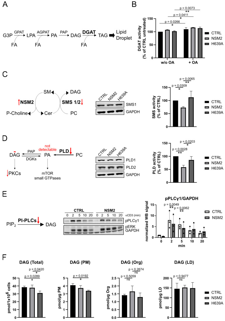
The accumulation of lipid droplets (LDs) and ceramides (Cer) is linked to non-alcoholic fatty liver disease (NAFLD), regularly co-existing with type 2 diabetes and decreased immune function. Chronic inflammation and increased disease severity in viral infections are the hallmarks of the obesity-related immunopathology. The upregulation of neutral sphingomyelinase-2 (NSM2) has shown to be associated with the pathology of obesity in tissues. Nevertheless, the role of sphingolipids and specifically of NSM2 in the regulation of immune cell response to a fatty acid (FA) rich environment is poorly studied. Here, we identified the presence of the LD marker protein perilipin 3 (PLIN3) in the intracellular nano-environment of NSM2 using the ascorbate peroxidase APEX2-catalyzed proximity-dependent biotin labeling method. In line with this, super-resolution structured illumination microscopy (SIM) shows NSM2 and PLIN3 co-localization in LD organelles in the presence of increased extracellular concentrations of oleic acid (OA). Furthermore, the association of enzymatically active NSM2 with isolated LDs correlates with increased Cer levels in these lipid storage organelles. NSM2 enzymatic activity is not required for NSM2 association with LDs, but negatively affects the LD numbers and cellular accumulation of long-chain unsaturated triacylglycerol (TAG) species. Concurrently, NSM2 expression promotes mitochondrial respiration and fatty acid oxidation (FAO) in response to increased OA levels, thereby shifting cells to a high energetic state. Importantly, endogenous NSM2 activity is crucial for primary human CD4+ T cell survival and proliferation in a FA rich environment. To conclude, our study shows a novel NSM2 intracellular localization to LDs and the role of enzymatically active NSM2 in metabolic response to enhanced FA concentrations in T cells.

Keywords: cholesteryl ester (CE); diacylglycerol (DAG); fatty acid oxidation (FAO); lipid droplet (LD); monounsaturated fatty acid (MUFA); neutral sphingomyelinase-2 (NSM2); plasma membrane (PM); triacylglycerol (TAG).


Conflict of interest statement

The authors declare no conflicts of interest. The funders had no role in the design of the study; in the collection, analyses, or interpretation of data; in the writing of the manuscript; or in the decision to publish the results.

Figures

Figure 1
Figure 1
NSM2-APEX2 proximity labeling identifies proteins related to exosome secretion and neutral lipid storage. (A) Workflow of proximity labeling strategy in living Jurkat cells expressing NSM2-APEX2. (B) Volcano plot for quantitative identification of the proteins proximal to NSM2. The x-axis indicates the logarithm-transformed ratio of the relative abundance of a protein in NSM2-APEX2 cells incubated with H2O2 versus cells where H2O2 was omitted (n = 3). The y-axis: the logarithm-transformed p-value of the Student’s test. Cut offs for differentially enriched proteins (relative abundance is changed more as 3.5-fold, negative log of p-value more as 3.6) are marked as red dotted lines. Each black dot represents one gene. LD associated protein PLIN3, LCP1 and TAGLN2 are highlighted in red. (C) Gene Ontology (GO) Biological Process analysis is shown for the 34 significantly enriched proteins within upper right quadrant in B. BP: biotin phenol; H2O2: hydrogen peroxide.
Figure 2
Figure 2
Intracellular NSM2 does not localize in the ER or organelles of the secretory pathway. (A) NSM (left graph) and ASM (right graph) activity in CTRL, NSM2 and H639A Jurkat cell lysates. (B) Cer (left column) and SM (right column) levels in lipid extracts of whole cells (upper graphs), PM (graphs in the middle) and organelle (Org) fractions (bottom graphs) of CTRL, NSM2 and H639A Jurkat cells as assessed using LC-MS/MS. (C) Western blot analysis of Org and PM fractions isolated from Jurkat cells. (D) Representative cropped fluorescence images of NSM2-GFP (green) Jurkat cells stained for different compartment markers (magenta) (left column, scale bar: 2 µm). Mander’s colocalization coefficients M1 for NSM2-GFP and each of the compartment markers are shown in the right column. Mean values with standard deviations of the measurements are shown (n = 3). p-values of one-way ANOVA with post hoc Turkey test are shown as * p < 0.05, ** p < 0.01, *** p < 0.001, and **** p < 0.0001; ns, non-significant.
Figure 3
Figure 3
Enzymatic activity is not required for NSM2 association with lipid droplets (LDs) in oleic acid (OA)-loaded cells. Representative SIM images of NSM2- and H639A-GFP Jurkat cells left untreated (A) or loaded with 300 µM OA overnight (B) and stained for PLIN3. Scale bar: 10 µm. Rectangles indicate the location of single cell zoom in shown below (scale bar: 2 µm). (C) Pearson correlation analysis for NSM2 or H639A co-localization with PLIN3 in cells incubated with OA (n = 68). (D) LD diameter in NSM2 and H639A cells shown in B (n = 87). (E) Representative Western blot (left) and densiometric quantification (right) of NSM2 and different cellular compartment markers in post nuclear supernatant (PNS) and LDs isolated from OA-treated cells (n = 3). Mean values with standard deviations of the measurements are shown. p-values of two-tailed student’s t-test analysis are shown as * p < 0.05 and **** p < 0.0001; ns, non-significant.
Figure 4
Figure 4
NSM2 activity in lipid droplets (LDs) does not affect their neutral lipid content. NSM activity and LC-MS lipid analysis of LDs isolated from CTRL, NSM2 and H639A cells treated with 300 µM OA overnight. (A) Quantification of NSM activity and sphingolipid (SM, Cer) content. (B) Levels of LD membrane lipids: MAG, PC, and neutral lipid: CE. (C) Levels of TAG species analyzed according to their acyl chain length (upper panels) or saturation (# of double bonds (DB); lower panels). Mean values with standard deviations of the measurements are shown (n = 3). p-values of one-way ANOVA with post hoc Turkey test are shown as * p < 0.05 and *** p < 0.001; ns, non-significant. (D) Heat maps of fold changes for different TAG species in LDs of CTRL vs. H639A (left) and LDs of CTRL vs. NSM2 (right). TAGs were plotted according to the number of carbons (rows) and double bonds (columns) of the FAs esterified to glycerol.
Figure 5
Figure 5
NSM2 activity impairs the cellular content of lipid droplets (LDs) and neutral lipids. (A) Analysis of CTRL, NSM2 and H639A cells treated with 300 µM OA overnight. Representative EM images (scale bar: 2 µm). Red arrows indicate LDs. Right graph: quantification of LD number per cell (n = 13). (B) Lipid analysis of TAG, PC and CE using LC-MS in total cell extracts (n = 3). (C) Quantification of TAG species according to their acyl chain length (upper panels) or saturation (# of double bonds (DB); lower panels). (D) Heat maps of fold changes for TAG species in CTRL vs. H639A Jurkat cells (top) and CTRL vs. NSM2 Jurkat cells (bottom). TAGs were plotted according to the number of carbons (rows) and double bonds (columns) of the FAs esterified to glycerol. Mean values with standard deviations of the measurements are shown. p-values of one-way ANOVA with post hoc Turkey test are shown as * p < 0.05, ** p < 0.01, *** p < 0.001, and **** p < 0.0001; ns, non-significant.
Figure 6
Figure 6
NSM2 activity negatively affects diycylglycerol (DAG) synthesis pathways upstream of neutral lipid triacylglycerol (TAG) accumulation. (A) Simplified scheme of the TAG synthesis pathway during LD biogenesis at the ER membrane. (B) Diacylglycerol acyltransferase (DGAT) activity of Jurkat cells loaded with 300 µM OA or left untreated was measured. (C) Sphingomyelin synthase (SMS) and (D) phospholipase D (PLD) enzymatic activities (right graphs) and protein levels (Western blot panels) in cell lysates. (E) Phosphorylation of phospholipase C-gamma 1 (PLCγ1) in cells stimulated with αCD3 for indicated time points. Red arrows indicate NSM2-dependent changes in enzymatic activity. (F) LC-MS measurements of diacylglycerol (DAG) levels in total cell extracts or subcellular fractions (PM, organelles (Org), LDs) of CTRL, NSM2 and H639A Jurkat cells. Mean values with standard deviations of three independent measurements are shown. p-values of one-way ANOVA with post hoc Turkey test are shown as * p < 0.05 and ** p < 0.01; ns, non-significant. DGK: DAG kinase; PIP2: phosphatidylinositol 4,5-bisphosphate; PI: phosphatidylinositol.
Figure 7
Figure 7
Increase in mitochondria size and fatty acid β-oxidation in cells expressing NSM2. Analysis of CTRL, NSM2 and H639A cells. (A) Representative fluorescence images (left panels) and quantification (right graph) of the mitochondrial footprints in individual cells (n = 107) labeled with MitoTracker™ Deep Red FM (scale bar: 20 µm). (B) Representative graphs of OCR in cells left untreated or treated overnight with OA (left). Quantification of maximal respiration and β-oxidation from three independent experiments (right graphs). Oligomycin (Oligo), FCCP, Etomoxir (Eto), Rotenone (Rot) and Antimycin A (AA) were injected during the measurements as indicated via the dotted lines. (C) Cellular energy phenotype profiles for OA-treated and untreated cells displayed as a scatter plot of OCR (y-axis) and ECAR (x-axis). Mean values with standard deviations of the measurements are shown in all graphs. p-values of one-way ANOVA with post hoc Turkey test (A) or two-tailed student’s t-test (B,C) are shown as * p < 0.05, ** p < 0.01, *** p < 0.001, and **** p < 0.0001; ns, non-significant.
Figure 8
Figure 8
Pharmacological inhibition of endogenous NSM2 enhances lipid droplet (LD) accumulation and cell death in primary CD4+ T cells treated with oleic acid (OA). Human CD4+ T cells were left untreated or pretreated with 1.5 µM ES048 for 2 h prior to co-stimulation with αCD3/αCD28 and loading with OA. (A) Representative fluorescence images of cells stained with BODIPY 493/503 after 3 days of treatment with 50 μM OA. Rectangles indicate the location of zoomed areas (right panels; scale bar: 20 µm). (B) Flow cytometry analysis of apoptotic (AnnexinV positive) and living (Annexin V and PI negative) cells (upper graph) and quantification of BODIPY 493/503 area per cell (n = 68) (lower graph) of CD4+ T cells shown in A. (C) Viability and (D) proliferation of CFSE labeled CD4+ T cells incubated with indicated concentrations of OA for 5 days and measured using flow cytometry. Right panel shows representative CFSE profiles of the cells loaded with 100 µM OA. (E) Analysis of maximal mitochondrial respiration and FA β-oxidation (left graphs, n = 3). Cellular energy phenotype profiles are displayed as a scatter plot of OCR (y-axis) and ECAR (x-axis) (right panel). Oligomycin (Oligo), FCCP, Etomoxir (Eto), Rotenone (Rot) and Antimycin A (AA) were injected during the measurements (n = 3). Mean values with standard deviations of the measurements are shown. p-values of two-way ANOVA with post hoc Sídák test are shown as * p < 0.05, ** p < 0.01, *** p < 0.001, and **** p < 0.0001; ns, non-significant.

Similar articles

Cited by

References

    1. Hartel J.C., Merz N., Groesch S. How sphingolipids affect T cells in the resolution of inflammation. Front. Pharmacol. 2022;13:1002915. doi: 10.3389/fphar.2022.1002915. - DOI - PMC - PubMed
    1. Kroll A., Cho H.E., Kang M.H. Antineoplastic Agents Targeting Sphingolipid Pathways. Front. Oncol. 2020;10:833. doi: 10.3389/fonc.2020.00833. - DOI - PMC - PubMed
    1. Avota E., de Lira M.N., Schneider-Schaulies S. Sphingomyelin Breakdown in T Cells: Role of Membrane Compartmentalization in T Cell Signaling and Interference by a Pathogen. Front. Cell Dev. Biol. 2019;7:152. doi: 10.3389/fcell.2019.00152. - DOI - PMC - PubMed
    1. Lee M., Lee S.Y., Bae Y.S. Functional roles of sphingolipids in immunity and their implication in disease. Exp. Mol. Med. 2023;55:1110–1130. doi: 10.1038/s12276-023-01018-9. - DOI - PMC - PubMed
    1. Hofmann K., Tomiuk S., Wolff G., Stoffel W. Cloning and characterization of the mammalian brain-specific, Mg2+-dependent neutral sphingomyelinase. Proc. Natl. Acad. Sci. USA. 2000;97:5895–5900. doi: 10.1073/pnas.97.11.5895. - DOI - PMC - PubMed