. 2024 Mar 15;25(6):3327.
doi: 10.3390/ijms25063327.

The DUX4-HIF1α Axis in Murine and Human Muscle Cells: A Link More Complex Than Expected

Affiliations

The DUX4-HIF1α Axis in Murine and Human Muscle Cells: A Link More Complex Than Expected

Thuy-Hang Nguyen et al. Int J Mol Sci. .

Abstract

FacioScapuloHumeral muscular Dystrophy (FSHD) is one of the most prevalent inherited muscle disorders and is linked to the inappropriate expression of the DUX4 transcription factor in skeletal muscles. The deregulated molecular network causing FSHD muscle dysfunction and pathology is not well understood. It has been shown that the hypoxia response factor HIF1α is critically disturbed in FSHD and has a major role in DUX4-induced cell death. In this study, we further explored the relationship between DUX4 and HIF1α. We found that the DUX4 and HIF1α link differed according to the stage of myogenic differentiation and was conserved between human and mouse muscle. Furthermore, we found that HIF1α knockdown in a mouse model of DUX4 local expression exacerbated DUX4-mediated muscle fibrosis. Our data indicate that the suggested role of HIF1α in DUX4 toxicity is complex and that targeting HIF1α might be challenging in the context of FSHD therapeutic approaches.

Keywords: DUX4; FSHD; HIF1α; myogenesis; skeletal muscle.


Conflict of interest statement

The authors declare no conflicts of interest.

Figures
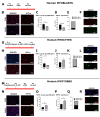
Figure 1
Figure 1
The differential effect of DUX4 on HIF1α expression and protein level in human LHCN-M2-iDUX4 muscle cells depends on the stage of differentiation. LHCN-M2-iDUX4 myoblasts were cultured and seeded as described in [40] at a standard PO2 of 21%. DUX4 expression was induced by the addition of 62.5 ng/mL of doxycycline (DOX) to the culture medium. For differentiation, cells at confluence were switched to a differentiation medium for two (myocytes) or four days (myotubes). Cells were fixed in 4% paraformaldehyde (PAF), and immunofluorescence (IF) was performed with antibodies directed against HIF1α or DUX4 and appropriate secondary antibodies coupled to Alexa Fluors. (A,G,M) Experiment time courses. (B,H,N) Representative fields showing HIF1α-positive (HIF1α+) nuclei (red IF). DAPI was used to visualize nuclei (blue). Scale bar = 100 µm. (C,I,O) Quantification of HIF1α+ nuclei normalized to the total number of nuclei (DAPI staining). Mean ± SEM, ** p < 0.01, t-test. (D,J,P) Relative HIF1A mRNA level normalized to RPLP0. Mean ± SEM, * p < 0.05, t-test. N = 4 for myoblasts, N = 3 for myocytes and myotubes. (E,K,Q) Proportion of DUX4+ nuclei among HIF1α+ nuclei. (F,L,R) Representative field showing HIF1α+ (red IF) and DUX4+ (green IF). Nuclei were stained with DAPI (blue). Scale bar = 50 µm. All experiments were performed on 3 independent cultures, each at least in triplicate. The total numbers of counted nuclei were, on average, 4719 for myoblasts, 5689 for myocytes and 24,486 for myotubes.
Figure 2
Figure 2
Effect of DUX4 induction on HIF1α target genes in human LHCN-M2-iDUX4 muscle cells. Cell culturing, induction of DUX4 expression by doxycycline and myogenic differentiation were performed at a standard PO2 of 21%, as in Figure 1. (A,E,I) Expression levels of PDK1 and VEGFA mRNAs. Quantifications were performed by RT-qPCR and normalized to RPLP0. Mean ± SEM, ** p < 0.01, *** p < 0.001, t-test. N = 4 for myoblasts, N = 3 for myocytes and myotubes. (B,F,J) PDK1 protein level determined by Western blot. Densitometry signal was normalized to total protein stained by Ponceau red. * p < 0.05, rank sum test, N = 6 for myoblasts and myocytes, N = 3 for myotubes. (C,G,K) Representative Western blot and Ponceau red staining for PDK1 detection. (D,H,L) VEGF protein level determined by ELISA on the culture medium. Mean ± SEM, * p < 0.05, t-test. N = 3.
Figure 3
Figure 3
Effect of DUX4 on the Hif1α pathway in murine myoblasts and myocytes. (AE) C2C12-iDUX4 murine myoblasts. A total of 25,000 or 200,000 cells were seeded per well in 24-well or 6-well plates, respectively, and grown at a standard PO2 of 21%. After 24 h, DUX4 expression was induced for 24 h with increasing doses of doxycycline (DOX, ng/mL). HIF1α was detected by immunofluorescence (IF). For comparison, LHCN-M2-iDUX4 myoblasts were cultured as in Figure 1, and DUX4 expression was induced for 24 h with increasing doses of DOX. (FJ) C2C12-iDUX4 murine myocytes. A total of 750,000 cells were seeded per well in 6-well plates. After 24 h, cells were switched to the differentiation medium for two days. DUX4 expression was induced with 62.5 ng/mL of DOX for 48 h. (A,F) Experiment time courses. (B,G) Representative fields showing Hif1α+ nuclei (red IF). Nuclei were stained with DAPI (blue). Scale bar = 100 µm. (C) Quantification of Hif1α+ nuclei normalized to the total number of nuclei (DAPI staining) in LHCN-M2-iDUX4 myoblasts and C2C12-iDUX4 myoblasts. Mean ± SEM, ** p < 0.01, *** p < 0.01, one-way ANOVA with Holm–Sidak post hoc test vs. the control (DOX: 0 ng/mL). (H) Quantification of HIF1α+ nuclei normalized to the total number of nuclei (DAPI staining) in C2C12- iDUX4 myocytes. Mean ± SEM, *** p < 0.001, t-test. (D,I) Expression level of Wfdc3 mRNA. Quantifications were performed by RT-qPCR and normalized to Rplp0. Mean ± SEM, * p < 0.05, *** p < 0.001, t-test. (E,J) Expression levels of Hif1a, Pdk1 and Vegfa mRNAs. Quantifications were performed by RT-qPCR and normalized to Rplp0. Mean ± SEM, * p < 0.05, t-test. Experiments were performed on 3 independent cultures, each in triplicate (N = 3). The total numbers of counted cells were on average 6809 for myoblasts and 3095 for myocytes.
Figure 4
Figure 4
Early effects of DUX4 expression on HIF1α pathway in vivo in the DUX4 IMEP (Intramuscular Electroporation) mouse model with a high dose of DUX4 expression. (A,C) Experiment time courses. (B) mRNAs of DUX4 target genes Wfdc3 and Zscan4c were quantified by RT-qPCR in C2C12 myoblasts 4, 5 and 6 h post transfection with pCIneo-DUX4. Quantifications were normalized to Rplp0. N = 5 for control group, and N = 4 for 4 h, 5 h and 6 h groups. Results are presented as boxplots. * p < 0.05. Kruskal–Wallis followed by Dunn’s post hoc test. (D) Representative sections of TA electroporated with saline solution (Left), 20 µg of pCIneo (Middle) or pCIneo-DUX4 plasmid (Right) stained using Hematoxylin–Eosin–Heindehain blue (HEB). Scale bar = 100 µm. (E) Effect of DUX4 induction on the level of Wfdc3 mRNA in the IMEP model. mRNA levels were quantified by RT-qPCR and normalized to Rplp0. Results are presented as boxplots. * p < 0.05. Kruskal–Wallis followed by Dunn’s post hoc test. N = 8 for each group. (F) Effect of DUX4 induction on the level of Hif1α pathway mRNAs Hif1a, Pdk1 and Vegfa in the IMEP model. mRNA levels were quantified by RT-qPCR and normalized to Rplp0. Results are presented as boxplots. * p < 0.05. Kruskal–Wallis followed by Dunn’s post hoc test. N = 8 for each group.
Figure 5
Figure 5
Involvement of Hif1α pathway in DUX4-induced muscle damage. (A) Efficiency of siRNA directed against Hif1a mRNA (siHIF1α). The TA muscle was electroporated with either saline solution, siCTL or siHIF1α. TA muscles were harvested 1 day after the IMEP procedure. Hif1a mRNA level was quantified by RT-qPCR and normalized to Rplp0. Mean ± SEM, * p < 0.05, *** p < 0.001. Kruskal–Wallis followed by Dunn’s post hoc test. N = 10 per group. (B) Representative cryosections of TA electroporated with 20 µg of pCIneo-DUX4 plasmid in combination or not with 2 µg of siCTL or siHIF1α. TA muscles were harvested 7 days after the IMEP procedure. Muscle sections were stained with HEB. Top: global view of the muscle sections; scale bar = 500 µm. Bottom: magnification of the damaged area; scale bar = 100 µm. (C) The percentage of lesion area was evaluated on muscle stained with HEB as represented in B. Data presented as scatter plots with mean ± SEM, * p < 0.05. One-way ANOVA followed by Holm–Sidak post hoc test. N = 8 per group. (D) Myofiber cross-section areas (CSA) were measured on the whole muscle section by using ImageJ software (https://imagej.net/ij/). Mean ± SEM, one-way ANOVA: NS. N = 8 per group. (E) Muscle fiber size distribution. Myofibers were classified in clusters according to their area. Chi-squared: NS. N = 8 per group. (F) Cumulative percentage of myofibers in clusters. (G) Wfdc3 mRNA level was quantified by RT-qPCR in the IMEP model. Quantifications were normalized to Rplp0. Data are presented as boxplots. Kruskal–Wallis followed by Dunn’s post hoc test: NS. N = 8 per group. (H) Effect of DUX4 induction on levels of Hif1α pathway mRNAs Hif1a, Pdk1 and Vegfa in the IMEP model. mRNA levels were quantified by RT-qPCR and normalized to Rplpl0. Data are presented as boxplots. Kruskal–Wallis followed by Dunn’s post hoc test: NS. N = 8 per group.
Figure 6
Figure 6
Schematic conclusions ❶ DUX4 inhibits HIF1α pathway in proliferating myoblasts but ❷ induces it in late differentiation into myotubes. Data regarding the two HIF1α target genes VEGFA and PDK1 are consistent with those results. However, ❸ DUX4 decreases PDK1 protein levels regardless of the differentiation stage, likely due to DUX4-induced mitochondrial dysfunction. ❹ A DUX4–HIF1α axis also exists in mouse myoblasts as well as is adult muscle in vivo. Moreover, as we described in [40] in the context of adult myogenesis, hypoxia ❺ increases early myogenic differentiation in a HIF1α-dependent way and ❻ induces myocyte fusion independently of HIF1α. Finally, ❼ DUX4 represses HIF1α’s effects in early myoblast differentiation.

References

    1. Banerji C.R.S., Zammit P.S. Pathomechanisms and Biomarkers in Facioscapulohumeral Muscular Dystrophy: Roles of DUX4 and PAX7. EMBO Mol. Med. 2021;13:e13695. doi: 10.15252/emmm.202013695. - DOI - PMC - PubMed
    1. Wang L.H., Tawil R. Facioscapulohumeral Dystrophy. Curr. Neurol. Neurosci. Rep. 2016;16:66. doi: 10.1007/s11910-016-0667-0. - DOI - PubMed
    1. Dixit M., Ansseau E., Tassin A., Winokur S., Shi R., Qian H., Sauvage S., Mattéotti C., van Acker A.M., Leo O., et al. DUX4, a Candidate Gene of Facioscapulohumeral Muscular Dystrophy, Encodes a Transcriptional Activator of PITX1. Proc. Natl. Acad. Sci. USA. 2007;104:18157–18162. doi: 10.1073/pnas.0708659104. - DOI - PMC - PubMed
    1. Lemmers R.J.L.F., van der Vliet P.J., Klooster R., Sacconi S., Camaño P., Dauwerse J.G., Snider L., Straasheijm K.R., van Ommen G.J., Padberg G.W., et al. A Unifying Genetic Model for Facioscapulohumeral Muscular Dystrophy. Science. 2010;329:1650–1653. doi: 10.1126/science.1189044. - DOI - PMC - PubMed
    1. Geng L.N., Yao Z., Snider L., Fong A.P., Cech J.N., Young J.M., van der Maarel S.M., Ruzzo W.L., Gentleman R.C., Tawil R., et al. DUX4 Activates Germline Genes, Retroelements and Immune-Mediators: Implications for Facioscapulohumeral Dystrophy. Dev. Cell. 2012;22:38–51. doi: 10.1016/j.devcel.2011.11.013. - DOI - PMC - PubMed

MeSH terms

LinkOut - more resources