. 2024 Mar 19;25(6):3431.
doi: 10.3390/ijms25063431.

Effect of Donor Age on Endocrine Function of and Immune Response to Ovarian Grafts

Affiliations

Effect of Donor Age on Endocrine Function of and Immune Response to Ovarian Grafts

Monica A Wall et al. Int J Mol Sci. .

Abstract

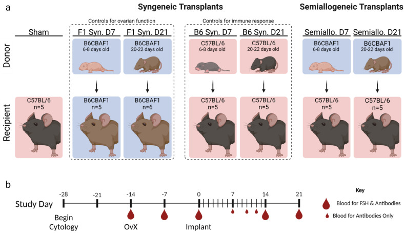
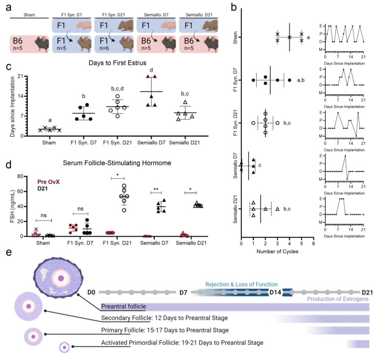
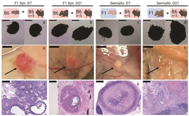
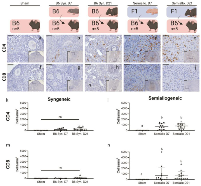
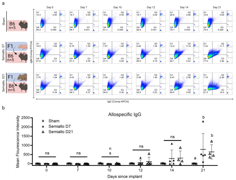
Premature loss of ovarian function (POI) is associated with numerous negative side effects, including vasomotor symptoms, sleep and mood disturbances, disrupted urinary function, and increased risks for osteoporosis and heart disease. Hormone replacement therapy (HRT), the standard of care for POI, delivers only a subset of ovarian hormones and fails to mimic the monthly cyclicity and daily pulsatility characteristic of healthy ovarian tissue in reproductive-aged individuals whose ovarian tissue contains thousands of ovarian follicles. Ovarian tissue allografts have the potential to serve as an alternative, cell-based HRT, capable of producing the full panel of ovarian hormones at physiologically relevant doses and intervals. However, the risks associated with systemic immune suppression (IS) required to prevent allograft rejection outweigh the potential benefits of comprehensive and dynamic hormone therapy. This work investigates whether the age of ovarian tissue donor animals affects the function of, and immune response to, subcutaneous ovarian grafts. We performed syngeneic and semi-allogeneic ovarian transplants using tissue from mice aged 6-8 (D7) or 20-22 (D21) days and evaluated ovarian endocrine function and immune response in a mouse model of POI. Our results revealed that tissue derived from D7 donors, containing an ample and homogeneous primordial follicle reserve, was more effective in fully restoring hypothalamic-pituitary-ovarian feedback. In contrast, tissue derived from D21 donors elicited anti-donor antibodies with higher avidity compared to tissue from younger donors, suggesting that greater immunogenicity may be a trade-off of using mature donors. This work contributes to our understanding of the criteria donor tissue must meet to effectively function as a cell-based HRT and explores the importance of donor age as a factor in ovarian allograft rejection.

Keywords: donor age; ovarian transplant; primary ovarian insufficiency.


Conflict of interest statement

The authors declare no conflicts of interest.

Figures

Figure 1
Figure 1
Experimental Design. (a) Ovarian tissue from mice, either 6–8 (D7 groups) or 20–22 days old (D21 groups), was transplanted subcutaneously into ovariectomized adult mice. To ensure the volume of transplanted tissue was equal across all subjects, D7 groups received two whole ovaries and D21 groups received two quarter ovaries. Syngeneic transplants were performed with both C57BL/6 (B6) mice (serving as controls for host immune response) and B6CBAF1 (F1) mice (serving as controls for ovarian endocrine function). For semi-allogeneic transplants, B6 hosts received ovarian tissue from F1 pups. Vaginal cytology was collected from 2 weeks before ovariectomy to 21 days after transplant. (b) Blood was collected at specified time points for analysis of serum follicle-stimulating hormone (FSH) and/or donor-specific Immunoglobulin G (IgG).
Figure 2
Figure 2
Restoration of endocrine function after ovarian transplantation in syngeneic (Syn) and semiallogeneic (semiallo) hosts (a) Schematic of groups for which ovarian function was compared. (b) Number of cycles and representative traces of estrus cycles following ovarian transplant (E = estrus, M = metestrus, D = diestrus, and P = proestrus). Rejection of ovarian tissue in semi-allogeneic groups resulted in fewer estrus cycles observed in these hosts. Bars indicate mean ± SD. Statistical significance was determined using the Kruskal–Wallace test and Dunn’s correction for multiple comparisons with p < 0.05, letters represent statistically significant differences, where groups that share a letter are not sta-tistically significantly different and groups that do not share a letter are statistically significantly different at a p value of at least 0.05 (c) Days elapsed between ovarian transplant and resumption of estrous cyclicity. Bars indicate median ± IQR and red points indicate mice that never entered estrus (censored subjects). Log-rank tests were performed on the Kaplan Meier Survival curves of each group with p < 0.05 used to determine statistical significance. (d) Serum FSH before ovariectomy (red) and 21 days after ovarian transplant (black). Only D7 syngeneic transplants were capable of suppressing FSH secretion to pre-OvX levels. Bars indicate mean ± SD. Statistical significance was determined using the Kruskal–Wallace test with ns representing p < 0.05, * representing 0.01 < p < 0.05, and ** representing 0.001 < p < 0.01. (e) Comparison of time required for follicles at different developmental stages to begin producing estrogens with time required for rejection or impaired graft function. D21 ovaries contain preantral follicles and are capable of secreting estrogens at the time of transplant. D7 ovaries contain only immature follicles and may not reach sufficient maturity to induce cyclicity before immune rejection and loss of tissue function.
Figure 3
Figure 3
Graft Morphology and Histology for syngeneic (Syn) and semiallogeneic (semiallo) transplants. (ad) Isolated ovaries prior to implantation. Ovarian tissue from mice 6–8 (D7)- or 20–22 (D21)-days old was transplanted subcutaneously into ovariectomized adult hosts. To match the volume of transplanted tissue across all groups, D7 groups received two whole D7 ovaries (a,c) and D21 groups received two quarter-D21 ovaries (b,d); scale bars = 500 μm. (e–h) Ovarian grafts after 21 days in vivo. Semi-allogeneic grafts (g,h) appear white and shrunken while syngeneic grafts (e,f) are pink and well-vascularized scale bars = 1 mm. (il) Hematoxylin and eosin staining of explanted ovarian grafts. Semi-allogeneic grafts (k,l) contain no healthy follicles, but showed dense nuclear staining consistent with immune cell infiltration and graft rejection. In contrast, syngeneic grafts (i,j) contain healthy follicles at various stages of development (Primordial follicle (dashed circle), primary follicle (*), secondary follicle (**), preantral follicle (***), antral follicle (****)) scale bars = 200 μm.
Figure 4
Figure 4
Presence of CD4+ and CD8+ cells at the graft site for syngeneic (Syn) and semiallogeneic (semiallo) transplants. (aj) Immunohistochemical staining of explanted ovarian grafts. Brown staining indicates cells positive for CD4 (ae) or CD8 (fj); scale bars = 50 μm, insets = 200 μm. (kn) Density of CD4 and CD8 positive cells at the graft site. Semi-allogeneic grafts had significantly increased densities of CD4+ (i) and CD8+ cells. (n) T cell comparison with native ovaries. This increased T cell density is consistent with cell-mediated rejection at the site of transplant. In contrast, neither CD4+ (k) nor CD8+ (m) T cell densities were significantly elevated in syngeneic grafts compared to the native ovary. Bars indicate mean ± SD. Statistical significance was determined using the Kruskal–Wallace test and Dunn’s correction for multiple comparisons with p < 0.05 considered significant, ns represents p > 0.05, letters represent statistically significant differences, where groups that share a letter are not statistically significantly different and groups that do not share a letter are statis-tically significantly different at a p value of at least 0.05.
Figure 5
Figure 5
Production of allospecific antibodies after ovarian transplantation to semiallogeneic (semiallo) hosts. Flow cytometry of donor thymocytes incubated with host serum was used to evaluate the production of donor-specific Immunoglobulin M (IgM) and Immunoglobulin G (IgG) after ovarian transplant. (a) Representative scatterplots for each group demonstrate an increase in allospecific antibodies in host serum over the 21 days following ovarian allotransplants; x-axis = (IgG), y-axis = (IgM). (b) Presence of IgG in host serum as measured by mean fluorescence intensity (MFI). Allospecific IgG was detectable in the serum of a subset of semi-allogeneic (semiallo) hosts as early as 12 days post-transplantation. Overall, 80% of D7 and 100% of D21 subjects who received semi-allogeneic implants mounted a detectable allospecific antibody by 21 days post-transplantation. The fluorescence intensity for each subject is normalized to the value recorded at D0. Bars indicate mean ± SD. Statistical significance was determined using the Kruskal–Wallace test and Dunn’s correction for multiple comparisons with p < 0.05 considered significant, ns represents p > 0.05, letters represent statistically significant differences, where groups that share a letter are not statistically significantly different and groups that do not share a letter are statis-tically significantly different at a p value of at least 0.05.

Similar articles

Cited by

References

    1. Donnez J., Dolmans M.M. Cryopreservation and transplantation of ovarian tissue. Clin. Obstet. Gynecol. 2010;53:787–796. doi: 10.1097/GRF.0b013e3181f97a55. - DOI - PubMed
    1. Sheshpari S., Shahnazi M., Mobarak H., Ahmadian S., Bedate A.M., Nariman-Saleh-Fam Z., Nouri M., Rahbarghazi R., Mahdipour M. Ovarian function and reproductive outcome after ovarian tissue transplantation: A systematic review. J. Transl. Med. 2019;17:396. doi: 10.1186/s12967-019-02149-2. - DOI - PMC - PubMed
    1. El Khoudary S.R., Aggarwal B., Beckie T.M., Hodis H.N., Johnson A.E., Langer R.D., Limacher M.C., Manson J.E., Stefanick M.L., Allison M.A., et al. Menopause Transition and Cardiovascular Disease Risk: Implications for Timing of Early Prevention: A Scientific Statement from the American Heart Association. Circulation. 2020;142:e506–e532. doi: 10.1161/CIR.0000000000000912. - DOI - PubMed
    1. Christin-Maitre S. Use of Hormone Replacement in Females with Endocrine Disorders. Horm. Res. Paediatr. 2017;87:215–223. doi: 10.1159/000457125. - DOI - PubMed
    1. Chakrabarti R., Chakrabarti R. Prescribing hormone replacement therapy: Key considerations for primary care physicians. Br. J. Gen. Pract. 2023;73:330–332. doi: 10.3399/bjgp23X733473. - DOI - PMC - PubMed