

Inositol-Exchange Activity in Human Primordial Placenta
- PMID: 38542419
- PMCID: PMC10970434
- DOI: 10.3390/ijms25063436
Inositol-Exchange Activity in Human Primordial Placenta
Abstract
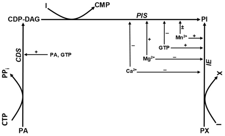
Human placenta is an intensively growing tissue. Phosphatidylinositol (PI) and its derivatives are part of the signaling pathway in the regulation of trophoblast cell differentiation. There are two different enzymes that take part in the direct PI synthesis: phosphatidylinositol synthase (PIS) and inositol exchange enzyme (IE). The presence of PIS is known in the human placenta, but IE activity has not been documented before. In our study, we describe the physiological properties of the two enzymes in vitro. PIS and IE were studied in different Mn2+ and Mg2+ concentrations that enabled us to separate the individual enzyme activities. Enzyme activity was measured by incorporation of 3[H]inositol in human primordial placenta tissue or microsomes. Optimal PIS activity was achieved between 0.5 and 2.0 mM Mn2+ concentration, but higher concentrations inhibit enzyme activity. In the presence of Mg2+, the enzyme activity increases continuously up to a concentration of 100 mM. PIS was inhibited by nucleoside di- and tri-phosphates. PI production increases between 0.1 and 10 mM Mn2+ concentration. The incorporation of [3H]inositol into PI increased by 57% when adding stabile GTP analog. The described novel pathway of inositol synthesis may provide an additional therapeutic approach of inositol supplementation before and during pregnancy.
Keywords: human placenta; inositol; phosphatidylinositol; phosphatidylinositol exchange enzyme; phosphatidylinositol synthase; placentation; trophoblast.
Conflict of interest statement
The authors declare no conflicts of interest.
Figures


Similar articles
-
Purification and characterization of phosphatidylinositol synthase from human placenta.Biochem J. 1994 Feb 1;297 ( Pt 3)(Pt 3):517-22. doi: 10.1042/bj2970517. Biochem J. 1994. PMID: 8110188 Free PMC article.
-
Phosphatidylinositol-inositol exchange in a rabbit lung.Biochim Biophys Acta. 1981 May 22;664(2):428-40. doi: 10.1016/0005-2760(81)90065-5. Biochim Biophys Acta. 1981. PMID: 6264967
-
Ubiquitous distribution of phosphatidylinositol phosphate synthase and archaetidylinositol phosphate synthase in Bacteria and Archaea, which contain inositol phospholipid.Biochem Biophys Res Commun. 2014 Jan 3;443(1):86-90. doi: 10.1016/j.bbrc.2013.11.054. Epub 2013 Nov 20. Biochem Biophys Res Commun. 2014. PMID: 24269814
-
Phosphatidylinositol synthase from yeast.Biochim Biophys Acta. 1997 Sep 4;1348(1-2):173-8. doi: 10.1016/s0005-2760(97)00103-3. Biochim Biophys Acta. 1997. PMID: 9370330 Review.
-
Phosphatidylinositol synthase from mammalian tissues.Biochim Biophys Acta. 1997 Sep 4;1348(1-2):179-86. doi: 10.1016/s0005-2760(97)00105-7. Biochim Biophys Acta. 1997. PMID: 9370331 Review.
References
-
- McKinnon T., Chakraborty C., Gleeson L.M., Chidiac P., Lala P.K. Stimulation of human extravillous trophoblast migration by IGF-II is mediated by IGF type 2 receptor involving inhibitory G protein(s) and phosphorylation of MAPK. J. Clin. Endocrinol. Metab. 2001;86:3665–3674. doi: 10.1210/jcem.86.8.7711. - DOI - PubMed
MeSH terms
Substances
LinkOut - more resources
Full Text Sources
Research Materials
Miscellaneous
