Aberrantly Glycosylated GLUT1 as a Poor Prognosis Marker in Aggressive Bladder Cancer
- PMID: 38542435
- PMCID: PMC10971045
- DOI: 10.3390/ijms25063462
Aberrantly Glycosylated GLUT1 as a Poor Prognosis Marker in Aggressive Bladder Cancer
Abstract
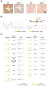
Muscle-invasive bladder cancer (MIBC) remains a pressing health concern due to conventional treatment failure and significant molecular heterogeneity, hampering the development of novel targeted therapeutics. In our quest for novel targetable markers, recent glycoproteomics and bioinformatics data have pinpointed (glucose transporter 1) GLUT1 as a potential biomarker due to its increased expression in tumours compared to healthy tissues. This study explores this hypothesis in more detail, with emphasis on GLUT1 glycosylation patterns and cancer specificity. Immunohistochemistry analysis across a diverse set of human bladder tumours representing all disease stages revealed increasing GLUT1 expression with lesion severity, extending to metastasis, while remaining undetectable in healthy urothelium. In line with this, GLUT1 emerged as a marker of reduced overall survival. Revisiting nanoLC-EThcD-MS/MS data targeting immature O-glycosylation on muscle-invasive tumours identified GLUT1 as a carrier of short glycosylation associated with invasive disease. Precise glycosite mapping uncovered significant heterogeneity between patient samples, but also common glycopatterns that could provide the molecular basis for targeted solutions. Immature O-glycosylation conferred cancer specificity to GLUT1, laying the molecular groundwork for enhanced targeted therapeutics in bladder cancer. Future studies should focus on a comprehensive mapping of GLUT1 glycosites for highly specific cancer-targeted therapy development for bladder cancer.
Keywords: GLUT1; bladder cancer; glycomics; glycoproteomics; targetable biomarkers.
Conflict of interest statement
André M.N. Silva, Lúcio Lara Santos and José Alexandre Ferreira are co-founders of GlycoMatters Biotech. The authors declare that this research was conducted in the absence of any commercial or financial relationships that could be construed as a potential conflict of interest. The funders had no role in the design of the study; in the collection, analyses, or interpretation of data; in the writing of the manuscript, or in the decision to publish the results.
Figures


Similar articles
-
Glycoproteomics identifies HOMER3 as a potentially targetable biomarker triggered by hypoxia and glucose deprivation in bladder cancer.J Exp Clin Cancer Res. 2021 Jun 9;40(1):191. doi: 10.1186/s13046-021-01988-6. J Exp Clin Cancer Res. 2021. PMID: 34108014 Free PMC article.
-
Target Score-A Proteomics Data Selection Tool Applied to Esophageal Cancer Identifies GLUT1-Sialyl Tn Glycoforms as Biomarkers of Cancer Aggressiveness.Int J Mol Sci. 2021 Feb 7;22(4):1664. doi: 10.3390/ijms22041664. Int J Mol Sci. 2021. PMID: 33562270 Free PMC article.
-
Targeted O-glycoproteomics explored increased sialylation and identified MUC16 as a poor prognosis biomarker in advanced-stage bladder tumours.Mol Oncol. 2017 Aug;11(8):895-912. doi: 10.1002/1878-0261.12035. Epub 2017 Mar 2. Mol Oncol. 2017. PMID: 28156048 Free PMC article.
-
[Recent advances in glycopeptide enrichment and mass spectrometry data interpretation approaches for glycoproteomics analyses].Se Pu. 2021 Oct;39(10):1045-1054. doi: 10.3724/SP.J.1123.2021.06011. Se Pu. 2021. PMID: 34505426 Free PMC article. Review. Chinese.
-
The clinicopathologic impacts and prognostic significance of GLUT1 expression in patients with lung cancer: A meta-analysis.Gene. 2019 Mar 20;689:76-83. doi: 10.1016/j.gene.2018.12.006. Epub 2018 Dec 12. Gene. 2019. PMID: 30552981 Review.
References
-
- Rosenberg J.E., Hoffman-Censits J., Powles T., van der Heijden M.S., Balar A.V., Necchi A., Dawson N., O’Donnell P.H., Balmanoukian A., Loriot Y., et al. Atezolizumab in patients with locally advanced and metastatic urothelial carcinoma who have progressed following treatment with platinum-based chemotherapy: A single-arm, multicentre, phase 2 trial. Lancet. 2016;387:1909–1920. doi: 10.1016/S0140-6736(16)00561-4. - DOI - PMC - PubMed
-
- Sharma P., Callahan M.K., Bono P., Kim J., Spiliopoulou P., Calvo E., Pillai R.N., Ott P.A., de Braud F., Morse M., et al. Nivolumab monotherapy in recurrent metastatic urothelial carcinoma (CheckMate 032): A multicentre, open-label, two-stage, multi-arm, phase 1/2 trial. Lancet Oncol. 2016;17:1590–1598. doi: 10.1016/S1470-2045(16)30496-X. - DOI - PMC - PubMed
MeSH terms
Substances
Grants and funding
LinkOut - more resources
Full Text Sources
Medical
Miscellaneous

