Review
. 2024 Mar 20;25(6):3520.
doi: 10.3390/ijms25063520.

Disruption of Neuromuscular Junction Following Spinal Cord Injury and Motor Neuron Diseases

Affiliations
Review

Disruption of Neuromuscular Junction Following Spinal Cord Injury and Motor Neuron Diseases

Colin Nemeth et al. Int J Mol Sci. .

Abstract

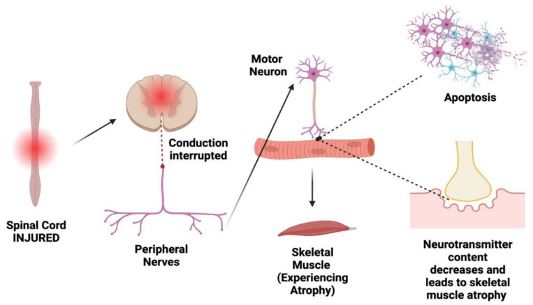
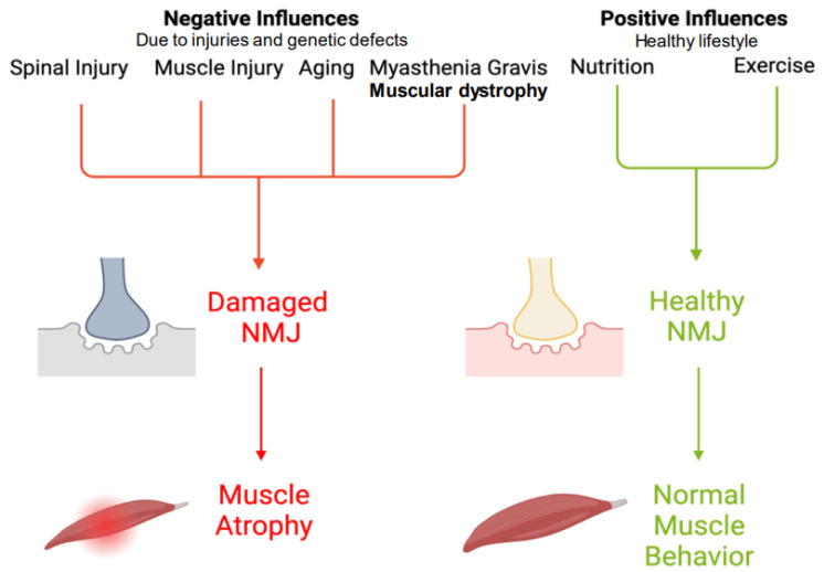
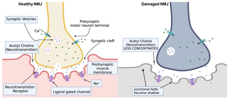
The neuromuscular junction (NMJ) is a crucial structure that connects the cholinergic motor neurons to the muscle fibers and allows for muscle contraction and movement. Despite the interruption of the supraspinal pathways that occurs in spinal cord injury (SCI), the NMJ, innervated by motor neurons below the injury site, has been found to remain intact. This highlights the importance of studying the NMJ in rodent models of various nervous system disorders, such as amyotrophic lateral sclerosis (ALS), Charcot-Marie-Tooth disease (CMT), spinal muscular atrophy (SMA), and spinal and bulbar muscular atrophy (SBMA). The NMJ is also involved in myasthenic disorders, such as myasthenia gravis (MG), and is vulnerable to neurotoxin damage. Thus, it is important to analyze the integrity of the NMJ in rodent models during the early stages of the disease, as this may allow for a better understanding of the condition and potential treatment options. The spinal cord also plays a crucial role in the functioning of the NMJ, as the junction relays information from the spinal cord to the muscle fibers, and the integrity of the NMJ could be disrupted by SCI. Therefore, it is vital to study SCI and muscle function when studying NMJ disorders. This review discusses the formation and function of the NMJ after SCI and potential interventions that may reverse or improve NMJ dysfunction, such as exercise, nutrition, and trophic factors.

Keywords: axonal damage; inflammation; myasthenia gravis; neuromuscular junction; skeletal muscle; spinal cord injury.


Conflict of interest statement

The authors declare no conflict of interest.

Figures

Figure 1
Figure 1
Relation of spinal cord injury to NMJ. Once a spinal cord experiences an injury, its signal conduction to the peripheral nerves is interrupted. Therefore, the motor neurons at the endplate experience cell death (apoptosis), the retransmitted content decreases, and, as a result, the skeletal muscle experiences atrophy. This sequence, starting with SCI, demonstrates how impactful the failure of nerve conduction can be in muscular function. Thus, SCI creates a non-functional NMJ that is hostile to motor neurons.
Figure 2
Figure 2
Negative and positive influences on NMJ. SCI as well as other neurodegenerative diseases (e.g., muscle injury, aging, and disorders such as muscular dystrophy and myasthenia gravis) facilitate damage to the NMJ, which therefore induces muscle atrophy. Nutrition and exercise are proactive measures to take that can avoid harming the NMJ. However, nutrition and exercise may not stop an SCI from harming the NMJ, but they may decrease the severity.
Figure 3
Figure 3
Healthy and functional NMJ compared to damaged NMJ due to SCI. Two enzymes (acetylcholinesterase and choline acetylase) play vital roles in maintaining NMJ integrity. A healthy NMJ has high neurotransmitter concentrations and regular junctional folds with neurotransmitter receptors. A damaged NMJ has limited concentrations of neurotransmitters and the junctional folds become shallow. After SCI, the motor neurons experience endplate death (apoptosis), making the NMJ damaged and dysfunctional in muscle use.

References

    1. Jones R.A., Harrison C., Eaton S.L., Llavero Hurtado M., Graham L.C., Alkhammash L., Oladiran O.A., Gale A., Lamont D.J., Simpson H., et al. Cellular and Molecular Anatomy of the Human Neuromuscular Junction. Cell Rep. 2017;21:2348–2356. doi: 10.1016/j.celrep.2017.11.008. - DOI - PMC - PubMed
    1. Jimsheleishvili S., Marwaha K., Sherman A.L. StatPearls. StatPearls Publishing; St. Petersburg, FL, USA: 2023. Physiology, Neuromuscular Transmission. - PubMed
    1. Hill M. The neuromuscular junction disorders. J. Neurol. Neurosurg. Psychiatry. 2003;74((Suppl. S2)):ii32–ii37. doi: 10.1136/jnnp.74.suppl_2.ii32. - DOI - PMC - PubMed
    1. Lee M., Kiernan M.C., Macefield V.G., Lee B.B., Lin C.S. Short-term peripheral nerve stimulation ameliorates axonal dysfunction after spinal cord injury. J. Neurophysiol. 2015;113:3209–3218. doi: 10.1152/jn.00839.2014. - DOI - PMC - PubMed
    1. Xu X., Talifu Z., Zhang C.J., Gao F., Ke H., Pan Y.Z., Gong H., Du H.Y., Yu Y., Jing Y.L., et al. Mechanism of skeletal muscle atrophy after spinal cord injury: A narrative review. Front. Nutr. 2023;10:1099143. doi: 10.3389/fnut.2023.1099143. - DOI - PMC - PubMed

MeSH terms