

Evaluating Fatty Acid Amide Hydrolase as a Suitable Target for Sleep Promotion in a Transgenic TauP301S Mouse Model of Neurodegeneration
- PMID: 38543105
- PMCID: PMC10975243
- DOI: 10.3390/ph17030319
Evaluating Fatty Acid Amide Hydrolase as a Suitable Target for Sleep Promotion in a Transgenic TauP301S Mouse Model of Neurodegeneration
Abstract
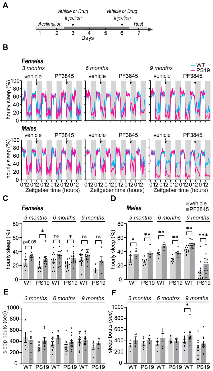
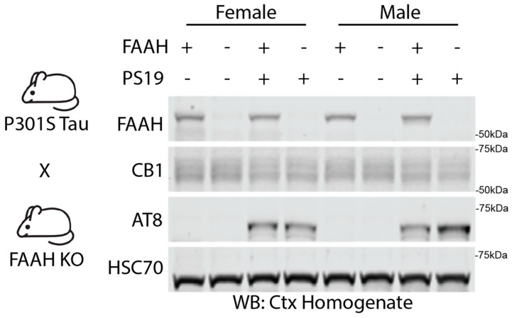
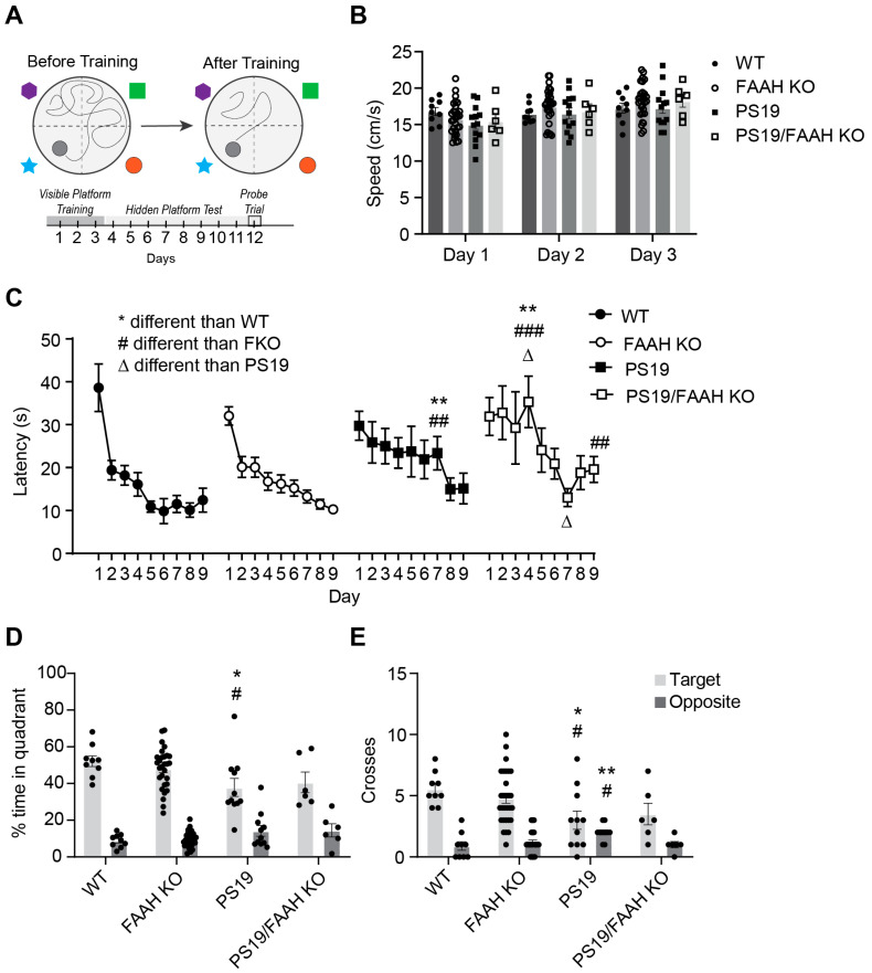
Sleep disruption is an expected component of aging and neurodegenerative conditions, including Alzheimer's disease (AD). Sleep disruption has been demonstrated as a driver of AD pathology and cognitive decline. Therefore, treatments designed to maintain sleep may be effective in slowing or halting AD progression. However, commonly used sleep aid medications are associated with an increased risk of AD, highlighting the need for sleep aids with novel mechanisms of action. The endocannabinoid system holds promise as a potentially effective and novel sleep-enhancing target. By using pharmacology and genetic knockout strategies, we evaluated fatty acid amide hydrolase (FAAH) as a therapeutic target to improve sleep and halt disease progression in a transgenic Tau P301S (PS19) model of Tauopathy and AD. We have recently shown that PS19 mice exhibit sleep disruption in the form of dark phase hyperarousal as an early symptom that precedes robust Tau pathology and cognitive decline. Acute FAAH inhibition with PF3845 resulted in immediate improvements in sleep behaviors in male and female PS19 mice, supporting FAAH as a potentially suitable sleep-promoting target. Moreover, sustained drug dosing for 5-10 days resulted in maintained improvements in sleep. To evaluate the effect of chronic FAAH inhibition as a possible therapeutic strategy, we generated FAAH-/- PS19 mice models. Counter to our expectations, FAAH knockout did not protect PS19 mice from progressive sleep loss, neuroinflammation, or cognitive decline. Our results provide support for FAAH as a novel target for sleep-promoting therapies but further indicate that the complete loss of FAAH activity may be detrimental.
Keywords: Alzheimer’s disease; FAAH; anandamide; endocannabinoids; sleep; tau.
Conflict of interest statement
The authors declare no conflicts of interest.
Figures


Similar articles
-
Sleep disruption precedes forebrain synaptic Tau burden and contributes to cognitive decline in a sex-dependent manner in the P301S Tau transgenic mouse model.bioRxiv [Preprint]. 2023 Jun 9:2023.06.07.544101. doi: 10.1101/2023.06.07.544101. bioRxiv. 2023. Update in: eNeuro. 2024 Jun 26;11(6):ENEURO.0004-24.2024. doi: 10.1523/ENEURO.0004-24.2024. PMID: 37333395 Free PMC article. Updated. Preprint.
-
Sleep Disruption Precedes Forebrain Synaptic Tau Burden and Contributes to Cognitive Decline in a Sex-Dependent Manner in the P301S Tau Transgenic Mouse Model.eNeuro. 2024 Jun 26;11(6):ENEURO.0004-24.2024. doi: 10.1523/ENEURO.0004-24.2024. Print 2024 Jun. eNeuro. 2024. PMID: 38858068 Free PMC article.
-
The role of endocannabinoid pathway in the neuropathology of Alzheimer's disease: Can the inhibitors of MAGL and FAAH prove to be potential therapeutic targets against the cognitive impairment associated with Alzheimer's disease?Brain Res Bull. 2021 Sep;174:305-322. doi: 10.1016/j.brainresbull.2021.06.022. Epub 2021 Jul 1. Brain Res Bull. 2021. PMID: 34217798 Review.
-
Potential Therapeutic Targets to Modulate the Endocannabinoid System in Alzheimer's Disease.Int J Mol Sci. 2024 Apr 5;25(7):4050. doi: 10.3390/ijms25074050. Int J Mol Sci. 2024. PMID: 38612861 Free PMC article. Review.
-
The role of fatty acid amide hydrolase enzyme inhibitors in Alzheimer's disease.Cell Biochem Funct. 2022 Mar;40(2):106-117. doi: 10.1002/cbf.3680. Epub 2021 Dec 21. Cell Biochem Funct. 2022. PMID: 34931308 Review.
Cited by
-
Fatty-acid amide hydrolase inhibition mitigates Alzheimer's disease progression in mouse models of amyloidosis.FEBS J. 2025 Aug;292(16):4160-4182. doi: 10.1111/febs.17403. Epub 2025 Jan 16. FEBS J. 2025. PMID: 39822137 Free PMC article.
-
Muscarinic Receptors and Alzheimer's Disease: New Perspectives and Mechanisms.Curr Issues Mol Biol. 2024 Jul 2;46(7):6820-6835. doi: 10.3390/cimb46070407. Curr Issues Mol Biol. 2024. PMID: 39057049 Free PMC article. Review.
References
-
- Martin S.C., Joyce K.K., Harper K.M., Nikolova V.D., Cohen T.J., Moy S.S., Diering G.H. Sleep disruption precedes forebrain synaptic Tau burden and contributes to cognitive decline in a sex-dependent manner in the P301S Tau transgenic mouse model. bioRxiv. 2023 doi: 10.1101/2023.06.07.544101. - DOI - PMC - PubMed
Grants and funding
LinkOut - more resources
Full Text Sources
