. 2024 Feb 29;12(3):501.
doi: 10.3390/microorganisms12030501.

A Pseudovirus-Based Neutralization Assay for SARS-CoV-2 Variants: A Rapid, Cost-Effective, BSL-2-Based High-Throughput Assay Useful for Vaccine Immunogenicity Evaluation

Affiliations

A Pseudovirus-Based Neutralization Assay for SARS-CoV-2 Variants: A Rapid, Cost-Effective, BSL-2-Based High-Throughput Assay Useful for Vaccine Immunogenicity Evaluation

Zhaohui Cai et al. Microorganisms. .

Abstract

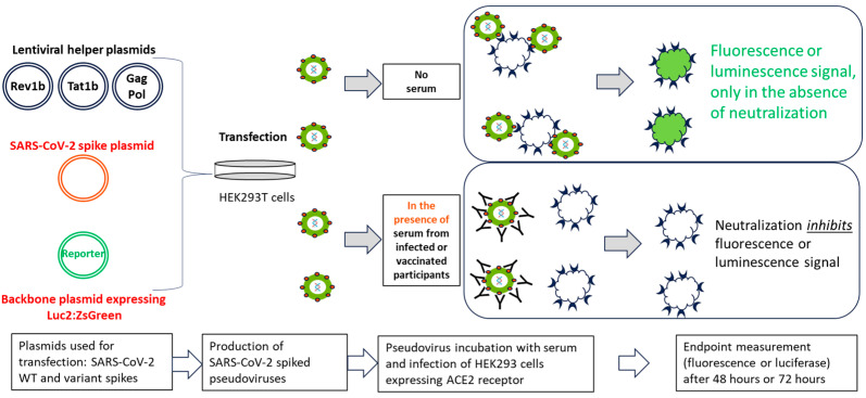
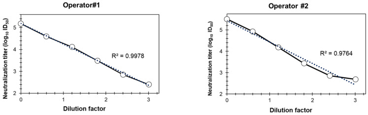
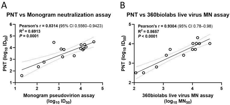
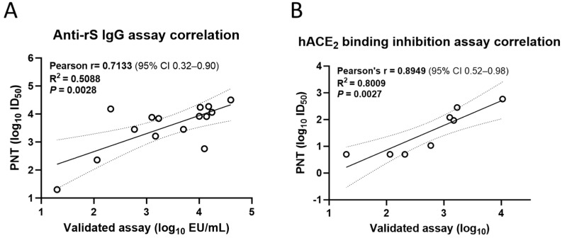
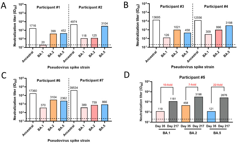
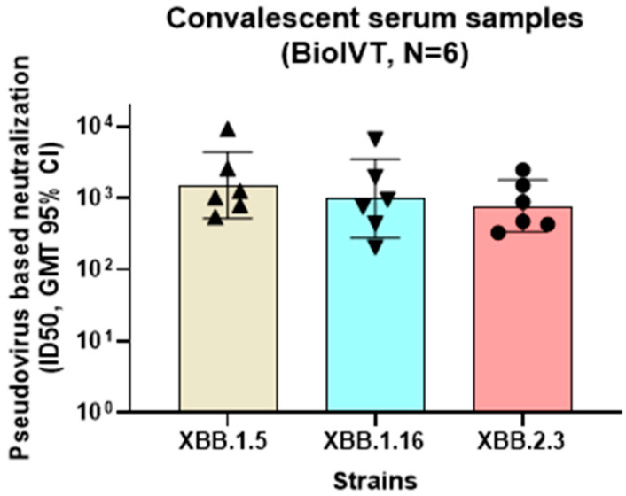
Neutralizing antibody responses from COVID-19 vaccines are pivotal in conferring protection against severe acute respiratory syndrome coronavirus 2 (SARS-CoV-2). Effective COVID-19 vaccines and assays measuring neutralizing antibodies against emerging variants (i.e., XBB.1.5, XBB.1.16, and XBB.2.3) are needed. The use of biosafety level (BSL)-3 laboratories for live virus assays results in higher costs and a longer turnaround time; therefore, a BSL-2-based pseudovirus neutralization assay (PNT) was developed. The pseudoviruses were produced by cotransfecting cells with plasmids encoding a lentiviral backbone-expressing luciferase reporter; non-surface proteins for lentiviral production; and ancestral or Omicron (BA.1 and BA.5) SARS-CoV-2 spike (S) proteins. The PNT was developed and optimized in dose and kinetics experiments. The representative serum samples (COVID-19-convalescent or NVX-CoV2373-vaccinated participants enrolled in the 2019nCoV-101 trial) demonstrated a wide dynamic range. The neutralization data showed robust correlation with validated anti-recombinant spike IgG levels and angiotensin-converting enzyme 2 inhibition titers (ancestral). This assay is suitable for measurement of the neutralization ability in clinical samples from individuals infected with SARS-CoV-2 or immunized with a COVID-19 vaccine. The results suggest that this PNT provides a lower cost, high-throughput, rapid turnaround alternative to BSL-3-based microneutralization assays and enables the discovery and development of effective vaccines against emerging variants.

Keywords: COVID-19; SARS-CoV-2; XBB.1.16; XBB.1.5; XBB.2.3; assay validation; correlate of protection; immunogenicity; neutralizing antibody titers; pseudovirus-based neutralization assays.


Conflict of interest statement

All the authors are employees and stockholders of Novavax, Inc. The authors declare that the research was conducted in the absence of any commercial or financial relationships that could be construed as a potential conflict of interest. Besides, the authors declare that this study received funding from Novavax, Inc. The funder was not involved in the study design, collection, analysis, interpretation of data, the writing of this article, or the decision to submit it for publication.

Figures

Figure 1
Figure 1
Schematic illustration of the PNT. Lentiviral helper plasmids (encoding Rev1b, Tat1b, Gag, and Pol proteins, lacking the gene for the envelope protein), a SARS-CoV-2 ancestral or variant S protein plasmid, and reporter genes (luc2 and ZsGreen) were cotransfected into HEK293T cells (adapted from Crawford, et al.) [13]. Transfected cell culture supernatants were used to infect 293T/hACE2 cells in the presence or absence of serum from infected or vaccinated participants, followed by measurement of the reporter protein expression (either luminescence or fluorescence). Neutralization of pseudovirus expressing SARS-CoV-2 S protein by the anti-S antibodies inhibits the expression of the reporter protein; therefore, the RLU reading in the presence of the test serum is inversely proportional to the neutralization index of that serum. ACE2, angiotensin-converting enzyme 2; HEK, human embryonic kidney; PNT, pseudovirus neutralization assay; SARS-CoV-2, severe acute respiratory syndrome coronavirus 2; WT, wild type.
Figure 2
Figure 2
PNT optimization: luciferase endpoint. RLU of 293T/hACE2 cells infected with different amounts of pseudoviruses (as represented by viral dilution factors on the X-axes) expressing either the S protein of SARS-CoV-2 (A) ancestral strain, (B) BA variants, or (C) XBB variants. RLU was measured 2 days (ancestral and Omicron variants) or 3 days (XBB variants) post infection. hACE2, human angiotensin-converting enzyme 2; RLU, relative luminescence units.
Figure 3
Figure 3
Suitability of cell lines for pseudovirus infections. (A) Three different cell lines (Vero-E6 (left), A549/hACE2-TMPRSS2 [overexpressing hACE2 and TMPRSS2] (middle), and 293T/hACE2 (right)) were infected with various amounts of pseudoviruses expressing either ancestral or variant (Omicron BA.1, BA.2, and BA.5) strain S proteins. RLU levels in the infected cells after 2 days are shown on the Y-axis. (B) Luminescence measurement of 293T/hACE2 and A549/hACE2-TMPRSS2 cells infected with Omicron BA.1 (left) or BA.5 (right) pseudoviruses in the presence or absence of increasing dilutions of serum (#2127). Cells infected with the pseudoviruses in the absence of test serum #2127 or cultured with no pseudoviruses were used as virus and cell controls, respectively. Percent neutralization compared to the virus control is shown on the Y-axis. hACE2, human angiotensin-converting enzyme 2; RLU, relative luminescence units; TMPRSS2, transmembrane serine protease 2.
Figure 4
Figure 4
Pseudovirus dose dependence and kinetics of endpoint (luminescence). (A) Four test serum samples were evaluated in the PNT, with increasing amounts of the ancestral strain pseudovirus (100, 250, and 500 TCID50/well). ID50 values of each serum are shown on the Y-axis. (B) Percent neutralization of serum dilutions with 250 TCID50/well of ancestral strain pseudovirus at each dilution level.
Figure 5
Figure 5
PNT precision. Human serum samples were evaluated in the PNT on different days. Duplicate samples were also tested in the same assay to evaluate intra-assay precision. Neutralization titers (ID50, GMT, 95% CI) are plotted on the Y-axis for the (A) ancestral, (B) Omicron BA.1, and (C) Omicron BA.5 strains. GMT, geometric mean titer; PNT, pseudovirus neutralization assay.
Figure 6
Figure 6
Linearity of the PNT. A test serum (#2127) was serially diluted (6 times, 4-fold dilution series), followed by evaluation with the ancestral strain, in duplicate at each dilution and by two operators, as described in Methods. Neutralization titers (log10 ID50) of the different dilutions are plotted on Y-axis. PNT, pseudovirus neutralization assay. Dotted line is the trend line and solid line is connectivity between the actual data points.
Figure 7
Figure 7
Correlation of the PNT with validated neutralization assays. Test serum samples from a clinical study were evaluated against the ancestral strain as per the method mentioned above, followed by regression analysis with a (A) validated neutralization assay and (B) live virus microneutralization assay (n = 13). Analysis was performed using GraphPad Prism® software (9.3.1). Dotted line depicts the 95% CI. Solid line is Pearson’s correlation line. MN, microneutralization; PNT, pseudovirus neutralization assay.
Figure 8
Figure 8
Correlation of the PNT with validated immunological assays. Test serum samples from a clinical study were evaluated in the PNT against the ancestral strain as per the method mentioned above, followed by regression analysis with a (A) validated anti-rS IgG assay (n = 15) and (B) a validated hACE2 binding inhibition assay (n = 8) Analysis was performed using GraphPad Prism® software (9.3.1). Dotted line shows 95% CI. Solid line is Pearson’s correlation line. hACE2, human angiotensin-converting enzyme 2; IgG, immunoglobulin G; PNT, pseudovirus neutralization assay; rS, recombinant spike; S, spike.
Figure 9
Figure 9
Clinical utility of the PNT. (AC) Serum samples were used from participants #1 to 4, 6, and 7 from a clinical study where they were vaccinated with NVX-CoV2373. Samples were tested in the PNT against the ancestral or a variant (Omicron BA.1, BA.2, and BA.5) strain. (D) Neutralization titers (ID50) for serum from participant #5, collected on day 35 (14 days after primary series vaccination with NVX-CoV2373) and day 217 (28 days after a booster dose). The horizontal dashed line represents the limit of detection. PNT, pseudovirus neutralization assay.
Figure 10
Figure 10
Neutralization activity of convalescent serum samples against Omicron XBB strains. Convalescent serum samples (BioIVT) were evaluated for neutralization activity in the PNT for XBB strains (XBB.1.5, XBB.1.16, and XBB.2.3). Serum samples showing >500 titer ID50 (n = 6) against XBB.1.5 were compared for neutralization activity against XBB.1.16 and XBB.2.3. No significant differences were observed (ordinary one-way ANOVA analysis, multiple comparisons (p > 0.5). GMT, geometric mean titer; PNT, pseudovirus neutralization assay.

Similar articles

Cited by

References

    1. Khoury D.S., Cromer D., Reynaldi A., Schlub T.E., Wheatley A.K., Juno J.A., Subbarao K., Kent S.J., Triccas J.A., Davenport M.P. Neutralizing antibody levels are highly predictive of immune protection from symptomatic SARS-CoV-2 infection. Nat. Med. 2021;27:1205–1211. doi: 10.1038/s41591-021-01377-8. - DOI - PubMed
    1. Abebe E.C., Dejenie T.A. Protective roles and protective mechanisms of neutralizing antibodies against SARS-CoV-2 infection and their potential clinical implications. Front. Immunol. 2023;14:1055457. doi: 10.3389/fimmu.2023.1055457. - DOI - PMC - PubMed
    1. Fong Y., Huang Y., Benkeser D., Carpp L.N., Áñez G., Woo W., McGarry A., Dunkle L.M., Cho I., Houchens C.R., et al. Immune correlates analysis of the PREVENT-19 COVID-19 vaccine efficacy clinical trial. Nat. Commun. 2023;14:331. doi: 10.1038/s41467-022-35768-3. - DOI - PMC - PubMed
    1. World Health Organization Tracking SARS-CoV-2 variants. [(accessed on 31 January 2024)]. Available online: https://www.who.int/activities/tracking-SARS-CoV-2-variants.
    1. Stanford University Coronavirus Antiviral and Resistance Database SARS-CoV-2 Variants. [(accessed on 31 January 2024)]. Available online: https://covdb.stanford.edu/variants/omicron_xbb/

LinkOut - more resources