. 2024 Feb 27;16(3):363.
doi: 10.3390/v16030363.

Discovery of a Novel Antiviral Effect of the Restriction Factor SPOC1 against Human Cytomegalovirus

Affiliations

Discovery of a Novel Antiviral Effect of the Restriction Factor SPOC1 against Human Cytomegalovirus

Anna K Kuderna et al. Viruses. .

Abstract

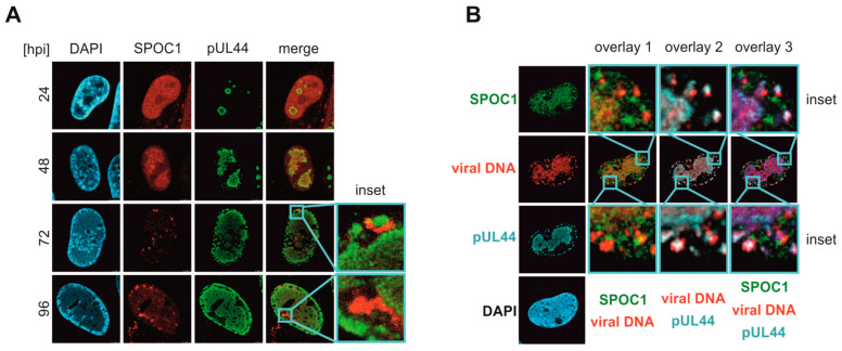
The chromatin-remodeler SPOC1 (PHF13) is a transcriptional co-regulator and has been identified as a restriction factor against various viruses, including human cytomegalovirus (HCMV). For HCMV, SPOC1 was shown to block the onset of immediate-early (IE) gene expression under low multiplicities of infection (MOI). Here, we demonstrate that SPOC1-mediated restriction of IE expression is neutralized by increasing viral titers. Interestingly, our study reveals that SPOC1 exerts an additional antiviral function beyond the IE phase of HCMV replication. Expression of SPOC1 under conditions of high MOI resulted in severely impaired viral DNA replication and viral particle release, which may be attributed to inefficient viral transcription. With the use of click chemistry, the localization of viral DNA was investigated at late time points after infection. Intriguingly, we detected a co-localization of SPOC1, RNA polymerase II S5P and polycomb repressor complex 2 (PRC2) components in close proximity to viral DNA in areas that are hypothesized to harbor viral transcription sites. We further identified the N-terminal domain of SPOC1 to be responsible for interaction with EZH2, a subunit of the PRC2 complex. With this study, we report a novel and potent antiviral function of SPOC1 against HCMV that is efficient even with unrestricted IE gene expression.

Keywords: RNA Pol II; antiviral; chromatin remodeler; dual role; human cytomegalovirus; restriction factor; viral transcription.


Conflict of interest statement

The authors declare no conflicts of interest.

Figures

Figure 1
Figure 1
Increasing HCMV doses are able to antagonize SPOC1-mediated repression of IE1 and IE2 expression. (A) The 24 hpi lysates of HCMV-infected control fibroblasts (HFF/Ctrl) and fibroblasts expressing SPOC1 (HFF/SPOC1) infected with HCMV TB40/E at MOIs of 0.01 (lanes 1 and 2), 1 (lanes 3 and 4) and 3 (lanes 5 and 6) were investigated by Western blotting. Expression levels of viral immediate-early proteins IE1 and IE2, β-actin and SPOC1 were analyzed. (B) Quantification of IE1 and IE2 signal intensities normalized to β-actin levels in HFF/SPOC1 relative to normalized IE1 and IE2 levels in HFF/Ctrl of three independent experiments. Statistical analysis was performed using Student’s t-test (one sample, two-tailed); **** p < 0.0001, * p < 0.05, n.s. = not significant.
Figure 2
Figure 2
SPOC1 expression negatively affects HCMV DNA replication and viral particle release. HFF/Ctrl and HFF/SPOC1 were infected in triplicate with HCMV strain TB40/E at an MOI of 1 or 3. (A) At 96 hpi, supernatants were analyzed for viral genome equivalents via qPCR. (B) At 96 hpi, intracellular DNA was isolated and HCMV genome equivalents were quantified via qPCR and normalized to albumin copy numbers. One out of three independent experiments is shown. Statistical analysis was performed using Student’s t-test (unpaired, two-tailed); *** p < 0.001, **** p < 0.0001. (C) Experimental set-up: Doxycycline (Dox)-inducible HFF/SPOC1 cells were either treated with Dox 24 h prior to or 24 h post infection with AD169 at MOI 0.1. (D) Dox-inducible HFF/SPOC1 was infected with AD169, at MOI 0.1, in triplicate. The infected cells were either left untreated or treated with Dox 24 h prior to or 24 h post infection (C). At 96 hpi, the supernatant was harvested and analyzed for viral genome equivalents via qPCR. One out of two experiments is shown. Statistical analysis was performed utilizing the one sample t-test; * p < 0.05.
Figure 3
Figure 3
SPOC1 leads to lower expression levels of viral early and late proteins. (A) Lysates of mock-infected (m) or TB40/E- (MOI of 3) or AD169-infected (MOI of 3 and 1) HFF/Ctrl and HFF/SPOC1 cells were analyzed at 24 to 72 hpi by separation on a 10 % polyacrylamide gel followed by Western blot detection of indicated proteins. Expression kinetics of viral immediate-early protein IE1, viral early protein pUL44 and viral late proteins pp28 and MCP were investigated. Asterisks (*) highlight the protein bands that are attenuated upon SPOC1 expression. (B) Quantification of IE1 levels in HFF/SPOC1 normalized to β-actin are depicted as fold change of the normalized IE1 level of HFF/Ctrl (indicated by dashed line at y = 1). (C) Quantification of early and late viral proteins of HFF/SPOC1 normalized to β-actin are depicted as fold change of the regarding normalized protein levels of HFF/Ctrl (indicated by dashed line at y = 1).
Figure 4
Figure 4
Impaired transcription of viral early and late genes in HFF/SPOC1 cells at late times in the replicative cycle. HFF/Ctrl and HFF/SPOC1 cells were infected with AD169 at an MOI of 1. (A) The 24 hpi IE1 and IE2 protein levels were analyzed via SDS PAGE and Western blotting. The relative intensity values were normalized to β-actin. Quantification of three independent experiments is shown. Statistical analysis was performed using Student’s t-test (one sample, two-tailed); n.s. = not significant. (B) IE1, IE2 and US3 transcript levels normalized to GAPDH were evaluated at 8 hpi using qPCR. Shown are the mean values of triplicates of one out of two experiments. Statistical analysis was performed using Student’s t-test (one sample, two-tailed). (C) At 48 and 72 hpi, total cellular RNA was isolated, cDNA was synthesized and viral mRNA levels of two immediate-early, early and late genes were quantified via qPCR, respectively. Shown are the mean values of triplicates of one out of three experiments. Statistical analysis was performed with Student’s t-test (unpaired, two-tailed); ** p < 0.01, *** p < 0.001, **** p < 0.0001.
Figure 5
Figure 5
SPOC1 localization during the time course of HCMV infection. (A) HFF/mCherry-SPOC1 cells were infected with AD169 at MOI 1 and fixed at the indicated time points. An antibody against pUL44 was used in combination with the secondary antibody Alexa-488. DAPI staining was used to visualize the nucleus. (B) At 72 hpi, AD169-infected HFF/SPOC1 (MOI of 1) was treated with F-Ara-EdU prior to fixation at 96 hpi. Samples were stained for SPOC1 (secondary antibody: Alexa-488) as well as with an antibody against pUL44 in combination with the Alexa-647 antibody. Click chemistry was performed to visualize viral DNA (modified from [15]).
Figure 6
Figure 6
SPOC1 co-localizes with PRC2 components close to viral DNA. HFF/SPOC1 was infected with AD169 at an MOI of 1. At 96 hpi, the cells were fixed and treated with antibodies directed against SPOC1, pUL44 and either (A) EZH2 or (B) SUZ12. As secondary antibodies, Alexa-488 (SPOC1) and a combination of either mouse or rabbit Alexa-555 and mouse or rabbit Alexa-647 were used. DAPI signals visualize the nucleus (modified from [15]). (C,D) At 72 hpi, EdC was added to SPOC1-expressing cells prior to fixation at 96 hpi. The same antibodies against EZH2 (C) or SUZ12 (D) were used as for (A) and (B); viral DNA was visualized by click chemistry. Nuclei were visualized by DAPI staining. Merge images were created from the two images above.
Figure 7
Figure 7
Interaction between SPOC1 deletion mutants and EZH2. (A) Schematic representation of the generated FLAG-SPOC1 deletion mutants with indicated amino acid sequence. (B) HEK293T cells were co-transfected with an empty control plasmid or FLAG-SPOC1 deletion mutants together with an EZH2-expressing plasmid. FLAG-SPOC1 was precipitated, and lysate controls as well as immuno-precipitated (IP) samples were analyzed via Western blotting. SPOC1 was visualized using an anti-FLAG antibody. CoIP = Co-Immunoprecipitaton.
Figure 8
Figure 8
Co-localization of SPOC1 with RNA pol II S5P. HFF/SPOC1 was infected with AD169 (MOI 1) and fixed at 96 hpi. Indirect immunostaining was performed using antibodies against SPOC1 and (A) 4H8 antibody detecting specifically Ser5 phosphorylated RNA pol II (modified from [15]) or (B) 8WG16 antibody to mark general RNA Pol II localization. DAPI was used to stain cell nuclei.

References

    1. Schilling E.M., Scherer M., Stamminger T. Intrinsic Immune Mechanisms Restricting Human Cytomegalovirus Replication. Viruses. 2021;13:179. doi: 10.3390/v13020179. - DOI - PMC - PubMed
    1. Schreiner S., Kinkley S., Bürck C., Mund A., Wimmer P., Schubert T., Groitl P., Will H., Dobner T. SPOC1-mediated antiviral host cell response is antagonized early in human adenovirus type 5 infection. PLoS Pathog. 2013;9:e1003775. doi: 10.1371/journal.ppat.1003775. - DOI - PMC - PubMed
    1. Hofmann S., Dehn S., Businger R., Bolduan S., Schneider M., Debyser Z., Brack-Werner R., Schindler M. Dual role of the chromatin-binding factor PHF13 in the pre- and post-integration phases of HIV-1 replication. Open Biol. 2017;7:170115. doi: 10.1098/rsob.170115. - DOI - PMC - PubMed
    1. Reichel A., Stilp A.C., Scherer M., Reuter N., Lukassen S., Kasmapour B., Schreiner S., Cicin-Sain L., Winterpacht A., Stamminger T. Chromatin-Remodeling Factor SPOC1 Acts as a Cellular Restriction Factor against Human Cytomegalovirus by Repressing the Major Immediate Early Promoter. J. Virol. 2018;92 doi: 10.1128/JVI.00342-18. - DOI - PMC - PubMed
    1. Mohrmann G., Hengstler J.G., Hofmann T.G., Endele S.U., Lee B., Stelzer C., Zabel B., Brieger J., Hasenclever D., Tanner B., et al. SPOC1, a novel PHD-finger protein: Association with residual disease and survival in ovarian cancer. Int. J. Cancer. 2005;116:547–554. doi: 10.1002/ijc.20912. - DOI - PubMed

LinkOut - more resources