Review
. 2024 Mar 12;16(3):442.
doi: 10.3390/v16030442.

Natural Adeno-Associated Virus Serotypes and Engineered Adeno-Associated Virus Capsid Variants: Tropism Differences and Mechanistic Insights

Affiliations
Review

Natural Adeno-Associated Virus Serotypes and Engineered Adeno-Associated Virus Capsid Variants: Tropism Differences and Mechanistic Insights

Estrella Lopez-Gordo et al. Viruses. .

Erratum in

Abstract

Today, adeno-associated virus (AAV)-based vectors are arguably the most promising in vivo gene delivery vehicles for durable therapeutic gene expression. Advances in molecular engineering, high-throughput screening platforms, and computational techniques have resulted in a toolbox of capsid variants with enhanced performance over parental serotypes. Despite their considerable promise and emerging clinical success, there are still obstacles hindering their broader use, including limited transduction capabilities, tissue/cell type-specific tropism and penetration into tissues through anatomical barriers, off-target tissue biodistribution, intracellular degradation, immune recognition, and a lack of translatability from preclinical models to clinical settings. Here, we first describe the transduction mechanisms of natural AAV serotypes and explore the current understanding of the systemic and cellular hurdles to efficient transduction. We then outline progress in developing designer AAV capsid variants, highlighting the seminal discoveries of variants which can transduce the central nervous system upon systemic administration, and, to a lesser extent, discuss the targeting of the peripheral nervous system, eye, ear, lung, liver, heart, and skeletal muscle, emphasizing their tissue and cell specificity and translational promise. In particular, we dive deeper into the molecular mechanisms behind their enhanced properties, with a focus on their engagement with host cell receptors previously inaccessible to natural AAV serotypes. Finally, we summarize the main findings of our review and discuss future directions.

Keywords: AAV engineering; AAV receptor; CNS; brain-blood barrier; directed evolution.


Conflict of interest statement

Thomas Weber is an employee of Spark Therapeutics. The other authors declare no conflicts of interest.

Figures

Figure 1
Figure 1
(A) Adeno-associated virus serotype (AAV) particle with the AAV single-stranded genome. (B) AAV2 genome organization including two inverted terminal repeats (ITs) flanking the rep and cap genes, cap polyA, and AAV promoters (p5, p19, and p40). (C) Transcriptional map of AAV structural (VP1, VP2, and VP3) and non-structural proteins (Rep78, Rep68, Rep52, Rep40, AAP, and MAAP). VP: viral protein; AAP: assembly-activating protein; and MAAP: membrane-associated AAV protein. Images created with Biorender (available at: https://www.biorender.com).
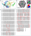
Figure 2
Figure 2
(A) Ribbon diagram of the AAV9 viral protein monomer colored by chain and with labelled β-sheet strands, α helix, VRs, and C/N-terminus. VR location within protein chains: VR-I (blue), VR-II (teal), VR-III (dark blue), VR-IV (green), VR-V and VR-VI (yellow), VR-VII (dark yellow), VR-VIII (orange), and VR-IX (red). (B) Adeno-associated virus (AAV) capsid surface model illustrates the location of variable regions (VR)-IV, V, and VIII (left panel). Representation of the AAV9 capsid zoomed-in (right panels), showing VR-IV at AA452-460 (red) and VR-VIII at AA581-593 (green) from a monomer and VR-V at AA488-505 (blue) from the adjacent monomer of the three-fold axis. (C) Comparison of VP1 amino acid sequences from AAV serotypes 1, 2, 3, 4, 6, 7, 8, and 9. VRs [43] are highlighted in red boxes. Multi-sequence alignment in (C) performed with Clustal Omega (available at: https://www.ebi.ac.uk/Tools/msa/clustalo/). Images in (A,B) constructed with PyMOL Molecular Graphics System Version 2.5.5, Schrodinger, LLC (Boston, MA, USA).
Figure 3
Figure 3
Model of entry and intracellular trafficking of adeno-associated virus (AAV) vectors. Following interactions with receptors and co-receptors and with the help of essential host cell components such as AAVR (1), AAV enters the target cell by means of endocytosis (2) and travels inside an endocytic vesicle toward the trans-Golgi network via retrograde trafficking (3). Acidification of the endosome and, potentially, additional factors trigger major structural changes within the capsid that expose a phospholipase A2 (PLA2) catalytic domain, allowing the virion to escape into the cytosol (4). The AAV virion can then be ubiquitinated for degradation (5a) or be imported into the nucleus through the nuclear pore complex (5b). In the nucleus, it accumulates in the nucleolus before moving into the nucleoplasm, where the single-stranded genome is released (6) and converted into a double-stranded DNA which can persist in the nucleus as a circular episome or as linear or episomal concatemers (7), leading to gene expression (8) and protein production (9). Image created with Biorender (available at https://www.biorender.com).
Figure 4
Figure 4
Location of anatomical structures of the human (A) and mouse (B) central nervous systems (CNSs). (C) Depiction of the brain vascular system with a zoomed-in cross-section of a vessel and the cell types composing it or adjacent to it. (D) Predominant cell types of the CNS. Images created with Biorender (available at https://www.biorender.com).
Figure 5
Figure 5
Schematic representing the genome arrangement of the library constructs of various AAV capsid engineering platforms. Images created with Biorender (available at https://www.biorender.com).

References

    1. Bedbrook C.N., Deverman B.E., Gradinaru V. Viral Strategies for Targeting the Central and Peripheral Nervous Systems. Annu. Rev. Neurosci. 2018;41:323–348. doi: 10.1146/annurev-neuro-080317-062048. - DOI - PubMed
    1. Buning H., Srivastava A. Capsid Modifications for Targeting and Improving the Efficacy of AAV Vectors. Mol. Ther. Methods Clin. Dev. 2019;12:248–265. doi: 10.1016/j.omtm.2019.01.008. - DOI - PMC - PubMed
    1. Campos L.J., Arokiaraj C.M., Chuapoco M.R., Chen X., Goeden N., Gradinaru V., Fox A.S. Advances in AAV technology for delivering genetically encoded cargo to the nonhuman primate nervous system. Curr. Res. Neurobiol. 2023;4:100086. doi: 10.1016/j.crneur.2023.100086. - DOI - PMC - PubMed
    1. Challis R.C., Ravindra Kumar S., Chen X., Goertsen D., Coughlin G.M., Hori A.M., Chuapoco M.R., Otis T.S., Miles T.F., Gradinaru V. Adeno-Associated Virus Toolkit to Target Diverse Brain Cells. Annu. Rev. Neurosci. 2022;45:447–469. doi: 10.1146/annurev-neuro-111020-100834. - DOI - PubMed
    1. Li C., Samulski R.J. Engineering adeno-associated virus vectors for gene therapy. Nat. Rev. Genet. 2020;21:255–272. doi: 10.1038/s41576-019-0205-4. - DOI - PubMed

Substances

LinkOut - more resources