. 2024 Mar 13;16(3):447.
doi: 10.3390/v16030447.

Immune System Deficiencies Do Not Alter SARS-CoV-2 Evolutionary Rate but Favour the Emergence of Mutations by Extending Viral Persistence

Affiliations

Immune System Deficiencies Do Not Alter SARS-CoV-2 Evolutionary Rate but Favour the Emergence of Mutations by Extending Viral Persistence

Laura Manuto et al. Viruses. .

Abstract

During the COVID-19 pandemic, immunosuppressed patients showed prolonged SARS-CoV-2 infections, with several studies reporting the accumulation of mutations in the viral genome. The weakened immune system present in these individuals, along with the effect of antiviral therapies, are thought to create a favourable environment for intra-host viral evolution and have been linked to the emergence of new viral variants which strongly challenged containment measures and some therapeutic treatments. To assess whether impaired immunity could lead to the increased instability of viral genomes, longitudinal nasopharyngeal swabs were collected from eight immunocompromised patients and fourteen non-immunocompromised subjects, all undergoing SARS-CoV-2 infection. Intra-host viral evolution was compared between the two groups through deep sequencing, exploiting a probe-based enrichment method to minimise the possibility of artefactual mutations commonly generated in amplicon-based methods, which heavily rely on PCR amplification. Although, as expected, immunocompromised patients experienced significantly longer infections, the acquisition of novel intra-host viral mutations was similar between the two groups. Moreover, a thorough analysis of viral quasispecies showed that the variability of viral populations in the two groups is comparable not only at the consensus level, but also when considering low-frequency mutations. This study suggests that a compromised immune system alone does not affect SARS-CoV-2 within-host genomic variability.

Keywords: SARS-CoV-2 genomic variability; immunocompromised subjects; intra-host; viral quasispecies.


Conflict of interest statement

The authors declare no conflicts of interest.

Figures

Figure 1
Figure 1
Infection length in immunocompromised and non-immunocompromised subjects. The figure reports the length of the SARS-CoV-2 infection in eight immunocompromised (Group 1) patients and in fourteen non-immunocompromised subjects at high risk of COVID-19 clinical progression (Group 2). The infection length is defined as the time window spanning from the first day of nasopharyngeal test positivity (day 0) to the first negative nasopharyngeal test. Group 1 subjects are reported in blue, whereas the different immunocompromised subjects are coloured according to the type of compromising condition, provided in the legend. The first negative test is presented as a grey dot. The other dots represent each day at which the subjects were tested, with filled dots indicating the availability of the viral sequence for that timepoint.
Figure 2
Figure 2
Emergence of SARS-CoV-2 consensus mutations in immunocompromised and non-immunocompromised subjects. The figure shows the longitudinal emergence of viral mutations in three immunocompromised (I) and in two non-immunocompromised (H) subjects. The longitudinal viral sequences of each patient are enclosed in a coloured box and are presented as coloured bars. Only the novel intra-host mutations compared to T0 are reported. Non-synonymous mutations are depicted as purple-filled diamonds, while silent mutations are presented as white diamonds. Deletions are indicated as black lines. Shaded mutations represent mutations present with a frequency below 50%.
Figure 3
Figure 3
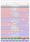
Deletions emerging in subject I_5 at T14. Visualisation of sequence reads spanning the SARS-CoV-2 genomic region 508–524 at T14 of the immunocompromised subject I_5. Reads mapping forward are reported in red, while reverse reads are depicted in blue. The black lines represent deletions, whose length is indicated in figures.
Figure 4
Figure 4
Quantitative analysis of minor variants. Comparison of the number of minor variants identified in immunocompromised (I) and in non-immunocompromised (H) subjects. (a) Comparison of the number of minor variants identified in immunocompromised and non-immunocompromised subjects, regardless of the timepoint at which the samples were collected (NI = 7, Mean I = 120.3, NH = 11, Mean H = 110. 0, p = 0.46, unpaired t test). (b) Comparison of the number of minor variants identified in immunocompromised and non-immunocompromised subjects at T0 (NI = 7, Mean I = 115.3, NH = 9, Mean H = 95.89, p = 0.10, unpaired t test). (c) Comparison of the number of minor variants identified in immunocompromised subjects and non-immunocompromised subjects at T7 (NI = 3, Mean I = 127.7, NH = 3, Mean H = 115.0, p = 0.49, unpaired t test). (d) Longitudinal intra-host variation of the number of minor variants identified in subjects for which T0 and T7 samples were available (NI = 3, NH = 2). The number of minor variants detected in each sample of an immunocompromised subjects are reported as red dots. Conversely, the number of minor variants detected in each sample of a non-immunocompromised subject are reported as blue dots, according to the legend.
Figure 5
Figure 5
Minor variant profiles of immunocompromised and non-immunocompromised subjects. (ac) Principal component analysis (PCA) of all the minor variants observed in samples collected from either the immunocompromised or the non-immunocompromised subjects at any timepoint (a); at T0 (b); and at T7 (c). According to the legend, minor variant profiles of immunocompromised subjects are provided as red dots, whereas the ones of non-immunocompromised subjects are presented as blue dots. Profiles of subjects infected with the Delta variant are represented as diamonds, whereas dots indicate Omicron as the variant of infection. The variation explained by each PC is provided along the relative axis. (de) Longitudinal analysis of the minor variants that persisted in all the considered timepoints (blue line), of the minor variants identified at least at one timepoint (red line) and of the minor variants specific to each specific timepoint in subject I_3 (d) and in subject I_5 (e).

References

    1. Choi B., Choudhary M.C., Regan J., Sparks J.A., Padera R.F., Qiu X., Solomon I.H., Kuo H.-H., Boucau J., Bowman K., et al. Persistence and Evolution of SARS-CoV-2 in an Immunocompromised Host. N. Engl. J. Med. 2020;383:2291–2293. doi: 10.1056/NEJMc2031364. - DOI - PMC - PubMed
    1. Beran A., Zink E., Mhanna M., Abugharbyeh A., Hanrahan J., Duggan J., Assaly R. Transmissibility and Viral Replication of SARS-CoV-2 in Immunocompromised Patients. J. Med. Virol. 2021;93:4156–4160. doi: 10.1002/jmv.26970. - DOI - PMC - PubMed
    1. Avanzato V.A., Matson M.J., Seifert S.N., Pryce R., Williamson B.N., Anzick S.L., Barbian K., Judson S.D., Fischer E.R., Martens C., et al. Case Study: Prolonged Infectious SARS-CoV-2 Shedding from an Asymptomatic Immunocompromised Individual with Cancer. Cell. 2020;183:1901–1912.e9. doi: 10.1016/j.cell.2020.10.049. - DOI - PMC - PubMed
    1. Quaranta E.G., Fusaro A., Giussani E., D’Amico V., Varotto M., Pagliari M., Giordani M.T., Zoppelletto M., Merola F., Antico A., et al. SARS-CoV-2 Intra-Host Evolution during Prolonged Infection in an Immunocompromised Patient. Int. J. Infect. Dis. 2022;122:444–448. doi: 10.1016/j.ijid.2022.06.023. - DOI - PMC - PubMed
    1. Kemp S.A., Collier D.A., Datir R.P., Ferreira I.A.T.M., Gayed S., Jahun A., Hosmillo M., Rees-Spear C., Mlcochova P., Lumb I.U., et al. SARS-CoV-2 Evolution during Treatment of Chronic Infection. Nature. 2021;592:277–282. doi: 10.1038/s41586-021-03291-y. - DOI - PMC - PubMed