. 2024 May;11(5):1250-1266.
doi: 10.1002/acn3.52041. Epub 2024 Mar 27.

Genome and RNA sequencing boost neuromuscular diagnoses to 62% from 34% with exome sequencing alone

Affiliations

Genome and RNA sequencing boost neuromuscular diagnoses to 62% from 34% with exome sequencing alone

Rhett G Marchant et al. Ann Clin Transl Neurol. 2024 May.

Abstract

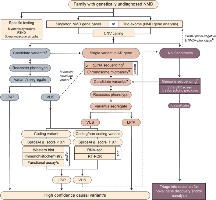
Objective: Most families with heritable neuromuscular disorders do not receive a molecular diagnosis. Here we evaluate diagnostic utility of exome, genome, RNA sequencing, and protein studies and provide evidence-based recommendations for their integration into practice.

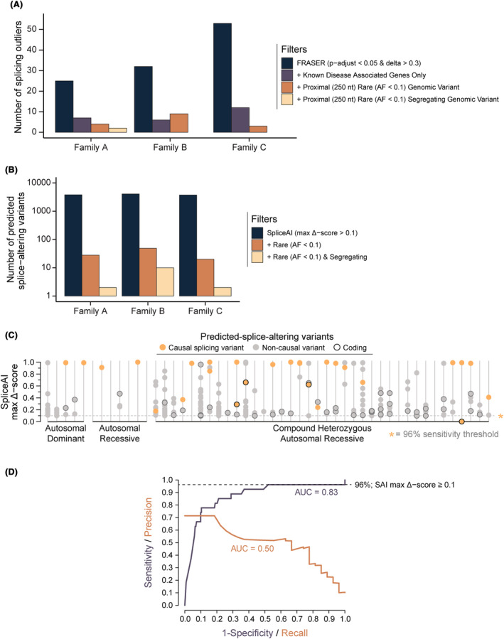
Methods: In total, 247 families with suspected monogenic neuromuscular disorders who remained without a genetic diagnosis after standard diagnostic investigations underwent research-led massively parallel sequencing: neuromuscular disorder gene panel, exome, genome, and/or RNA sequencing to identify causal variants. Protein and RNA studies were also deployed when required.

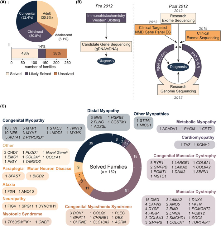
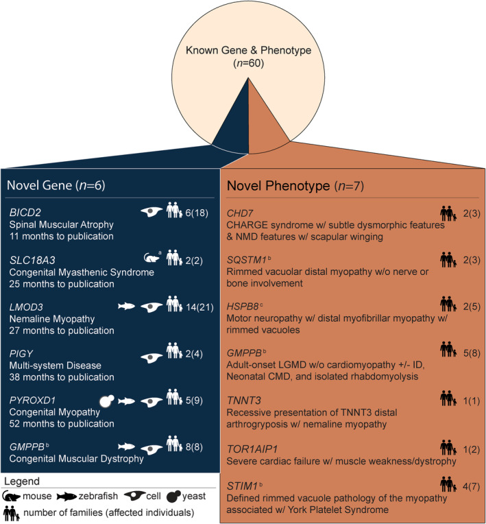
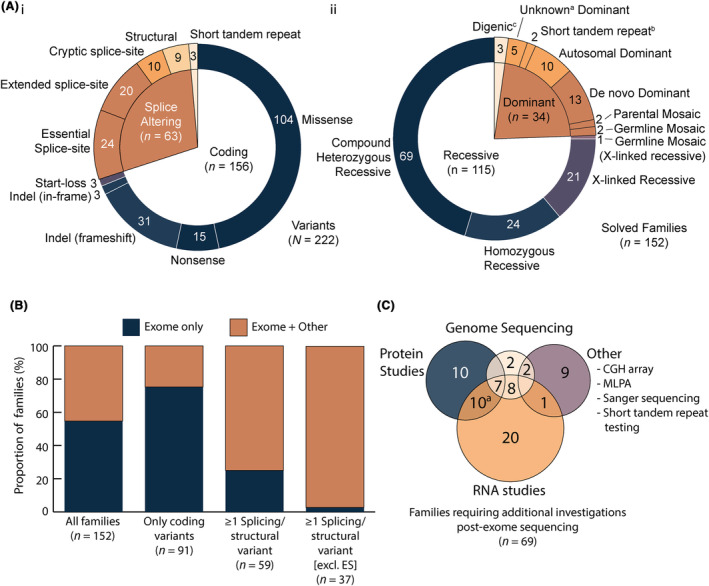
Results: Integration of exome sequencing and auxiliary genome, RNA and/or protein studies identified causal or likely causal variants in 62% (152 out of 247) of families. Exome sequencing alone informed 55% (83 out of 152) of diagnoses, with remaining diagnoses (45%; 69 out of 152) requiring genome sequencing, RNA and/or protein studies to identify variants and/or support pathogenicity. Arrestingly, novel disease genes accounted for <4% (6 out of 152) of diagnoses while 36.2% of solved families (55 out of 152) harbored at least one splice-altering or structural variant in a known neuromuscular disorder gene. We posit that contemporary neuromuscular disorder gene-panel sequencing could likely provide 66% (100 out of 152) of our diagnoses today.

Interpretation: Our results emphasize thorough clinical phenotyping to enable deep scrutiny of all rare genetic variation in phenotypically consistent genes. Post-exome auxiliary investigations extended our diagnostic yield by 81% overall (34-62%). We present a diagnostic algorithm that details deployment of genomic and auxiliary investigations to obtain these diagnoses today most effectively. We hope this provides a practical guide for clinicians as they gain greater access to clinical genome and transcriptome sequencing.


Conflict of interest statement

S.T. Cooper is director of Frontier Genomics Pty Ltd (Australia). S.T. Cooper receives no remuneration (salary or consultancy fees) for this role. Frontier Genomics Pty Ltd has no current financial interests that will benefit from publication of this data. S.T. Cooper is a named inventor on intellectual property owned jointly by the University of Sydney and Sydney Children's Hospitals Network. This IP relates to splicing variant detection and interpretation and is licensed by Frontier Genomics Pty Ltd. S.T. Cooper is named inventor on Australian Patent No. 2019379868 and Australian Provisional Patent No. 2019900836. The remaining co‐authors declare no competing interests.

Figures

Figure 1
Figure 1
Overview of research‐led diagnostic NMD cohort. (A) Age of onset of the 247 probands in our neuromuscular disease (NMD) cohort (i), with two‐thirds presenting with congenital (<2 years; n = 80) or pediatric‐onset (2–12 years; n = 76) disorders. A majority of the cohort is considered either solved (48%) or likely solved (14%) (ii). (B) Schematic of NMD diagnostic pipeline employed by our laboratory prior to, and after, 2012 when massively parallel sequencing was adopted as a first‐line investigation. (C) Research‐led massively parallel sequencing revealed a diverse set of genetic etiologies within NMD sub‐groups. The number of families in our cohort linked to each causal gene is listed. Genes are divided into disease groups according to observed phenotype and classification in the gene table of neuromuscular disorders. aNote, it is the collective opinion of our multidisciplinary team that digenic TP63 and DMPK variants are the likely causal basis for CMD in the proband. The proband is now deceased and there are no additional family members to gather further evidence supporting a digenic disorder. We therefore consider the family “likely solved” and are not pursuing further investigations. bA high confidence candidate novel disease gene in an advanced stage of functional validation. Three other genes that form part of the same complex are existing OMIM genes with a related phenotype. ES, exome sequencing; IHC, immunohistochemistry; WB, western blot.
Figure 2
Figure 2
Novel disease genes and phenotypes identified in the NMD cohort. Six novel genes were identified from the cohort: BICD2, SLC18A3, LMOD3, PIGY, PYROXD1, and GMPPB. a SLC18A3 was published citing two previously reported mouse models., The average time to publication for novel genes was 2·5 years. Seven novel phenotypes in known neuromuscular disorder genes were reported from our cohort: CHD7, SQSTM1, HSPB8, STIM1, GMPPB, TNNT3, and TOR1AIP1. Families from our cohort with novel disease genes and phenotypes were reported either by our group, by other groupsb (timeline not available), or in collaborationc. CMD, congenital muscular dystrophy; GDD, global developmental delay; ID, intellectual disability; LGMD, limb girdle muscular dystrophy; NM, nemaline myopathy; NMD, neuromuscular disease.
Figure 3
Figure 3
Variants identified in the cohort by variant type, inheritance, and the investigations required to confirm them as causal. (A) The nature of different classes of pathogenic variants identified in our NMD cohort (i) and the pattern of inheritance (ii). Heterozygous variants for which segregation or family history was not available are labeled as unknown dominanta. bTrinucleotide repeat disorder testing was conducted in a diagnostic genetic laboratory. cNote the digenic inheritance count includes the unconfirmed TP63/DMPK family addressed in Figure 1. (B) Proportion of families diagnosed by exome sequencing alone or exome sequencing and additional investigations (+ Other) stratified by the variant types identified. (C) Venn diagram of additional investigations required for diagnosis post‐exome. aIncludes one case which underwent RNA studies, protein studies, and MLPA. AD, autosomal dominant; CGH, comparative genomic hybridization; ES, essential splice‐site; MLPA, multiplex ligation‐dependent probe amplification.
Figure 4
Figure 4
Trio genome sequencing data filters out highly scored but noncausal findings from in silico splicing analyses. (A) Deployment of FRASER to identify splicing outliers in 3 families with RNA‐Seq data available. Analyzing statistically significant splicing outliers identified by FRASER using the recommended thresholds (range 25–55 outliers per sample/case; adjusted p‐value <0.05 & effect‐size/delta‐psi ≥0.3) alongside genomic sequencing data was essential to reduce the large number of noncausal findings. In practice, by confirming the presence of a rare (allele frequency <0.1 and number of alternate allele homozygotes <5), segregating variant identified by genome sequencing proximal (±250 nt) to a splicing outlier all but two splicing outliers across the three families were excluded, substantially reducing the time required to manually validate candidate outliers (in Integrative Genomics Viewer 30 ). (B) Number of variants identified in genome sequencing data across all genes using SpliceAI delta score of ≥0.1 as threshold for a prediction of splice‐altering outcomes in the same three families as in S1a. Inclusion of trio segregation data reduced noncausal predicted‐splice‐altering variants by greater than 10‐fold. (C) Segregating rare variants from exome or genome data for 50 solved families with SpliceAI maximum delta scores ≥0.1. On average, families had 4.8 rare (allele frequency <0.1 & number of alternate allele homozygotes <5), noncausal predicted‐splice‐altering rare segregating variants in known NMD genes (n = 606). Searches were conducted in each family using the segregation of the known causal variant. For compound heterozygous recessive noncausal predicted‐splice‐altering variants, confirmation of a second rare variant in trans was not sought. Therefore, 4.8 noncausal predicted‐splice‐altering segregating variants per family is likely an overestimation. (D) Receiver‐operator characteristic curve showing a SpliceAI maximum delta score threshold of ≥0.1 results in a sensitivity of 96% for the detection of splice‐altering causal variants in the 50 solved families in S1c. Poor performance across the precision‐recall curve is indicative of the high number of noncausal segregating variants predicted to be splice‐altering by SpliceAI.
Figure 5
Figure 5
Proposed diagnostic algorithm for neuromuscular disorders. Our recommendation for genomic testing in neuromuscular disorders (NMD) begins with a high coverage, targeted‐capture NMD panel or exome sequencing interrogated for a regularly updated list of NMD disease genes. Depending on whether candidate variants are found in a phenotypically concordant genea, further resolution of VUSb or finding a second, likely‐splice altering or structural variant through gDNA sequencingc or microarrayd, may be necessary. When no candidate variants are detected from the initial screen, trio exome sequencing is only recommended when the presentation suggests other multi‐system abnormalities, with the potential for a causal gene not covered on an NMD panele. Instead, we recommend genome sequencing; singleton for X‐linked or consanguineous pedigrees, and trio genome for suspected autosomal recessive or de novo dominant pedigreesf. From either the panel, exome, or genome branches of the algorithm, variants are divided into suspected splice‐altering (SpliceAI delta score ≥0.1) or not (≤0.1) and functional studies conducted as detailed. We acknowledge that immunostaining, western blot, or functional assays to resolve pathogenicity of VUS are services not available to most tertiary centers. However, RNA testing is likely to become increasingly available over the next 5 years. Our previous work outlines clinically endorsed strategies for RNA testing. Due to the complexity of RNA and known caveats with alignment of short sequencing reads, we recommend confirming all reportable findings detected by RNA‐Seq by RT‐PCR. This algorithm provides a comprehensive interrogation of massively parallel sequencing data for coding, splicing, and structural variants in NMD. AR, autosomal recessive; CNV, copy number variant; FSHD, facioscapulohumeral muscular dystrophy; LP/P, likely pathogenic/pathogenic; NMD, neuromuscular disorder; RT‐PCR, reverse transcription PCR; STR, short tandem repeat; SV, structural variant; VUS, variant of uncertain significance.

References

    1. Laing NG. Genetics of neuromuscular disorders. Crit Rev Clin Lab Sci. 2012;49(2):33‐48. doi:10.3109/10408363.2012.658906 - DOI - PubMed
    1. Fraiman YS, Wojcik MH. The influence of social determinants of health on the genetic diagnostic odyssey: who remains undiagnosed, why, and to what effect? Pediatr Res. 2021;89(2):295‐300. doi:10.1038/s41390-020-01151-5 - DOI - PMC - PubMed
    1. Beecroft SJ, Lamont PJ, Edwards S, et al. The impact of next‐generation sequencing on the diagnosis, treatment, and prevention of hereditary neuromuscular disorders. Mol Diagn Ther. 2020;24(6):641‐652. doi:10.1007/s40291-020-00495-2 - DOI - PubMed
    1. North KN, Wang CH, Clarke N, et al. Approach to the diagnosis of congenital myopathies. Neuromuscul Disord. 2014;24(2):97‐116. doi:10.1016/j.nmd.2013.11.003 - DOI - PMC - PubMed
    1. Anderson LVB, Davison K. Multiplex Western blotting system for the analysis of muscular dystrophy proteins. Am J Pathol. 1999;154(4):1017‐1022. doi:10.1016/S0002-9440(10)65354-0 - DOI - PMC - PubMed