. 2024 Mar 13:15:1378989.
doi: 10.3389/fmicb.2024.1378989. eCollection 2024.

The alternative coproporphyrinogen III oxidase (CgoN) catalyzes the oxygen-independent conversion of coproporphyrinogen III into coproporphyrin III

Affiliations

The alternative coproporphyrinogen III oxidase (CgoN) catalyzes the oxygen-independent conversion of coproporphyrinogen III into coproporphyrin III

Toni Mingers et al. Front Microbiol. .

Abstract

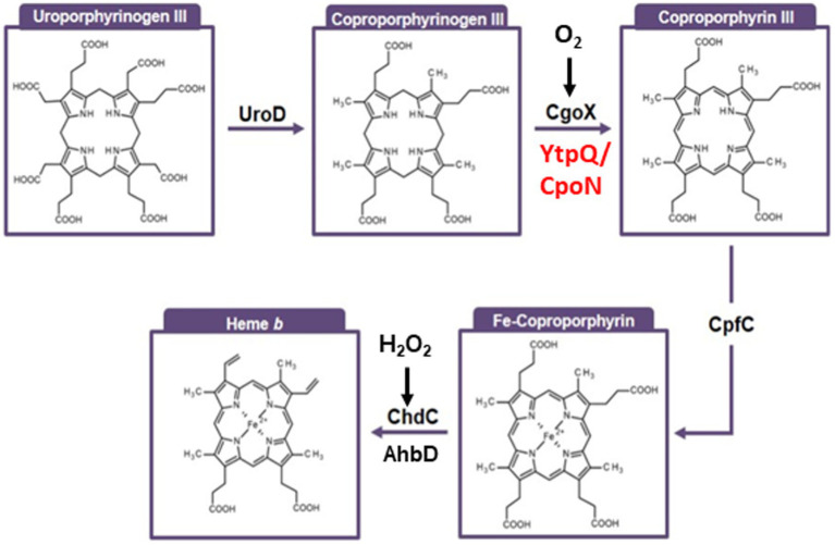
Nature utilizes three distinct pathways to synthesize the essential enzyme cofactor heme. The coproporphyrin III-dependent pathway, predominantly present in Bacillaceae, employs an oxygen-dependent coproporphyrinogen III oxidase (CgoX) that converts coproporphyrinogen III into coproporphyrin III. In this study, we report the bioinformatic-based identification of a gene called ytpQ, encoding a putative oxygen-independent counterpart, which we propose to term CgoN, from Priestia (Bacillus) megaterium. The recombinantly produced, purified, and monomeric YtpQ (CgoN) protein is shown to catalyze the oxygen-independent conversion of coproporphyrinogen III into coproporphyrin III. Minimal non-enzymatic conversion of coproporphyrinogen III was observed under the anaerobic test conditions employed in this study. FAD was identified as a cofactor, and menadione served as an artificial acceptor for the six abstracted electrons, with a KM value of 3.95 μmol/L and a kcat of 0.63 per min for the substrate. The resulting coproporphyrin III, in turn, acts as an effective substrate for the subsequent enzyme of the pathway, the coproporphyrin III ferrochelatase (CpfC). Under aerobic conditions, oxygen directly serves as an electron acceptor, but is replaced by the more efficient action of menadione. An AlphaFold2 model of the enzyme suggests that YtpQ adopts a compact triangular shape consisting of three domains. The N-terminal domain appears to be flexible with respect to the rest of the structure, potentially creating a ligand binding site that opens and closes during the catalytic cycle. A catalytic mechanism similar to the oxygen-independent protoporphyrinogen IX oxidase PgoH1 (HemG), based on the flavin-dependent abstraction of six electrons from coproporphyrinogen III and their potential quinone-dependent transfer to a membrane-localized electron transport chain, is proposed.

Keywords: Bacillaceae; Priestia megaterium; alternative heme biosynthesis; anaerobic metabolism; coproporphyrinogen III oxidase.


Conflict of interest statement

TM was employed by the company Pieris Pharmaceuticals GmbH. AC was employed by the company Syngenta UK Ltd. The remaining authors declare that the research was conducted in the absence of any commercial or financial relationships that could be construed as a potential conflict of interest. The author(s) declared that they were an editorial board member of Frontiers, at the time of submission. This had no impact on the peer review process and the final decision.

Figures

Figure 1
Figure 1
Pathway for the coproporphyrin-dependent heme b biosynthesis. This pathway is specific for Gram-positive Bacillaceae. First, decarboxylation catalyzed by UroD (uroporphyrinogen III decarboxylase) results in coproporphyrinogen III formation. Subsequent oxidation catalyzed by CgoX and CgoN (coproporphyrinogen III oxidases) leaves the name-giving coproporphyrin III intermediate. Next, iron is inserted by CpfC (coproporphyrin III ferrochelatase) to yield iron-coproporphyrin III (coproheme). A final decarboxylation is catalyzed by ChdC (coproheme decarboxylase) leading to the final product heme b.
Figure 2
Figure 2
Phylogenetic distribution of YtpQ and YptQ-like proteins across different bacteria. The phylogenetic tree was built using Mega 11-based maximum likelihood analyses with 500 bootstrap replicates for the YtpQ protein. The iTOL software was employed for tree display customization. Proteins belonging to Gram-negative bacterial strains are shown in blue. The query strain protein sequence is highlighted in red.
Figure 3
Figure 3
(A) SDS gel of the production and purification of P. megaterium YptQ (CgoN). Recombinant P. megaterium YptQ (CgoN) was produced in E. coli BL 21 as outlined in Material and Methods. The image shows an InstantBlue™ stained 12% SDS polyacrylamide gel after electrophoresis. Pierce™ unstained molecular weight marker protein ranging Mr from 14,000 to 116,000 are shown in lane 1. Further, total cellular extracts before IPTG induction (lane 2); 2 h (lane 3) and 20 h (lane 4) after the addition of 200 μM IPTG-are next; followed by the soluble (lane 5) and insoluble fraction of the extract (lane 6) after ultracentrifugation. The flow through of the glutathione agarose (lane 7), washing fraction 1 (lane 8); washing fraction 4 (lane 9), elution fraction 1 (lane 10) and fraction 2 (lane 11) after PreScission™ protease cleavage, and the final GST-tag elution fraction (lane 12) are concluding the analysis. Fraction 10 contains a single YptQ (CgoN) band with a Mr of 30,000 ± 3,000, nicely corresponding to the calculated molecular weight of the protein of 30,781 Da. (B) Native molecular weight and oligomerization state of P. megaterium YptQ determined via gel permeation chromatography. Analytical gel permeation chromatography of freshly purified recombinant P. megaterium YtpQ (CgoN) was performed using a 24 mL Superdex® 200 10/30 column on an Äkta purifier system under anaerobic conditions. Protein elution was followed by absorbance measurements at 280 nm. The column was equilibrated using the Gel Filtration Molecular Weight Markers Kit MWGF200 (Sigma Aldrich, Germany) composed of cytochrome c (Mr = 12,400), carbonic anhydrase (Mr = 29,000), bovine serum albumin (Mr = 66,000), alcohol dehydrogenase (Mr = 150,000), β-amylase (Mr = 200,000), and apoferritin (Mr = 400,000). Their elution position is given in Mr (x1,000) on the top of the figure. The chromatogram shows one major peak of Mr = 35,000 ± 5,000, indicating a monomeric YtpQ (CgoN) protein. (C) Time-resolved coproporphyrinogen III to coproporphyrin III conversion by P. megaterium YtpQ in the presence of FAD and menadione. Standard anaerobic YtpQ (CgoN) assays including 1 μM purified, recombinant YtpQ (CgoN), 10 μM coproporphyrinogen III, 3 μM FAD and 5 μM menadione were incubated at 30°C and 200 rpm. Samples were taken at different time points and analyzed by fluorescence spectroscopy with an excitation wavelength of 409 nm and emission measurements from 510 nm to 690 nm at 0 min (cyan), 5 min (turquoise), 10 min (yellow), 20 min (orange), 30 min (light blue), 45 min (dark blue), and 60 min (brown). (D) Kinetic analysis of coproporphyrinogen III oxidase activity of purified, recombinant P. megaterium YtpQ (CgoN). Anaerobic standard activity assays were conducted with 1 μM purified YtpQ (CgoN), 3 μM FAD and 5 μM menadione, and 1–20 μM of the substrate coproporphyrinogen III. A graphic representation of the initial velocity v0 of coproporphyrin III formation by the enzyme as a function of substrate concentration is shown. A KM value of 3.95 μmol/L and a kcat of 0.63 min−1 were deduced.
Figure 4
Figure 4
Mass spectrometric analysis and absorbance spectrum of the CgoN (YtpQ) product. Standard assays containing 1 μM freshly purified CgoN, 5 μM menadione, 3 μM FAD and 10 μM freshly prepared coproporphyrinogen III were at 30°C and 200 rpm for 120 min. Samples were analyzed after protein elimination via HPLC-MS. Shown are the HPLC run followed at 400 nm absorbance (upper panel), the spectroscopic analysis of the fraction causing the major absorbance peak in the upper panel (middle panel) and the corresponding mass spectrum. The product is coproporphyrin III with a typical absorbance maximum at 400 nm with side peaks between 480 nm and 600 nm and a molecular mass (m/z) of 655.
Figure 5
Figure 5
Conversion of YtpQ (CgoN)- derived coproporphyrin III by P. megaterium cf. into iron-coporporphyrin III (coproheme). In the first 300 μL assay 1 μM freshly purified YtpQ (CgoN), 5 μM menadione, 3 μM FAD, 10 μM freshly prepared coproporphyrinogen III were incubated at 30°C and 200 rpm for 120 min under anaerobic conditions (blue line). The second assay was composed identical to assay 1, but 1 μM freshly purified CpfC (HemH) and 10 μM (NH4)2Fe(SO4)2 were added additionally (red line). Both samples were HPLC-separated on a C18 column and the elution was followed by absorbance measurements at 400 nm. The column was calibrated with coproporphyrinogen III (27, 60 min) and Fe-coproporphyrinogen III (coproheme, 27, 75 min). The results of both experiments were combined in this figure. The addition of CpfC (HemH) and (NH4)2Fe(SO4)2 to YtpQ (CgoN) standard assay resulted in the formation of Fe-coproporphyrinogen III (coproheme).
Figure 6
Figure 6
Predicted structure of YtpQ from Priesta (Bacillus) megaterium DSM 319. (A) AlphaFold2 model obtained from the AlphaFold DB (entry D5DN20). The protein consists of 270 amino acids that fold into three domains (N-terminal domain: residues 1–78, blue; central domain: residues 79–161, yellow; C-terminal domain: residues 162–270, red). (B) Electrostatic potential mapped to the surface of the model shown in (A). Note the formation of a potential binding site at the interface between the N-terminal and central with C-terminal domains. Positive (blue) and negative (red) potential are displayed at ±100 KbT/ec. The electrostatic potential has been calculated with the APBS (Jurrus et al., 2018) plugin in PyMOL (Schiffrin et al., 2020).
Figure 7
Figure 7
Model of YtpQ (CgoN) activity. YtpQ (CgoN) catalyzes the oxidation of coproporphyrinogen III to coproporphyrin III transferring 6 electrons via its cofactor FAD to the quinone pool. Those electrons are further channeled into the redox systems of the various aerobic and anaerobic respiratory chains (simplified depiction as light blue square). Corresponding redox reactions cause the formation of a proton (alternatively sodium ion) gradient across the membrane that in turn fuels ATP synthesis via ATPase.

References

    1. Bali S., Lawrence A. D., Lobo S. A., Saraiva L. M., Golding B. T., Howard M. J., et al. . (2011). Molecular hijacking of siroheme for and d1 heme synthesis; elucidation of a new branch of tetrapyrrole synthesis. Proc. Natl. Acad. Sci. U. S. A. 108, 18260–18265. doi: 10.1073/pnas.1108228108, PMID: - DOI - PMC - PubMed
    1. Boynton T. O., Daugherty L. E., Dailey T. A., Dailey H. A. (2009). Identification of Escherichia coli HemG as a novel, menadione-dependent flavodoxin with protoporphyrinogen oxidase activity. Biochemistry 48, 6705–6711. doi: 10.1021/bi900850y, PMID: - DOI - PMC - PubMed
    1. Boynton T. O., Gerddes S., Craven H. H., Neidle E. L., Phillips J. D., Dailey H. A. (2011). Discovery of a gene involved in a third bacterial protoporphyrinogen oxidase activity through comparative genomic analysis and functional complementation. Appl Environ Microbiol. 77, 4795–801. doi: 10.1128/aem.00171-11 - DOI - PMC - PubMed
    1. Dailey T. A., Boynton T. O., Albetel A. N., Gerdes S., Johnson M. K., Dailey H. A. (2010). Discovery and characterization of HemQ: an essential heme biosynthetic pathway component. J. Biol. Chem. 285, 25978–25986. doi: 10.1074/jbc.M110.142604, PMID: - DOI - PMC - PubMed
    1. Dailey T. A., Dailey H. A. (1998). Identification of an FAD superfamily containing protoporphyrinogen oxidases, monoamine oxidases and phytoene desaturase. Expression and characterization of phytoene desaturase of Myxococcus xanthus. J. Biol. Chem. 273, 13658–13662. doi: 10.1074/jbc.273.22.13658, PMID: - DOI - PubMed

LinkOut - more resources