

Cavernous malformation located medially and deeply in the brain may be prone to false lateralization in cavernous malformation-associated epilepsy
- PMID: 38545148
- PMCID: PMC10966682
- DOI: 10.1016/j.heliyon.2024.e28273
Cavernous malformation located medially and deeply in the brain may be prone to false lateralization in cavernous malformation-associated epilepsy
Abstract
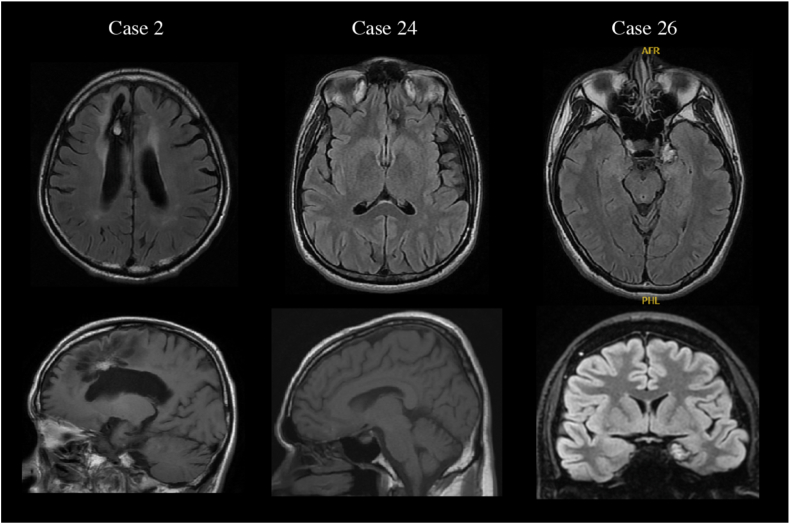
Background: Cavernous malformation (CM) is a well-known cause of epilepsy. Although the location of the CM is usually consistent with the side of seizure onset, some reports have described discrepancies between results from scalp electroencephalography (EEG) and CM location. This study investigated the prevalence and features of patients showing false lateralization (FL). Particularly, we tested the hypothesis that patients showing FL were more likely to have CM in medial and deep areas of the brain than in other areas.
Methods: Patients diagnosed with CM-associated epilepsy in our institution between March 2009 and March 2023 were included in this retrospective analysis. We investigated the presence or absence of FL of interictal epileptiform discharges (IEDs) or ictal discharges against MRI findings or against the true focus as determined from surgical outcomes. We compared the FL group with the non-false-lateralization group (NFL group) to clarify features of CM-associated epilepsy patients showing FL.
Results: Thirty-two epilepsy patients with CM were analyzed. The frequency of FL to MRI was 10.3% for IEDs and 7.7% for ictal discharges, while the frequency of FL to true focus after removal surgery was 10.5% for IEDs and 7.7% for ictal discharges. Regarding the FL of IEDs against MRI findings, the percentage of medial and deep lesions was significantly higher in the FL group (3/3, 100%) than in the NFL group (6/26, 23.1%; p = 0.023). No significant differences in age, sex, seizure type, or size of the CM were seen between groups.
Conclusions: CM-associated epilepsy can also present with FL, particularly if the location of the CM is medial and deep. Caution may be needed in determining the area for resection based solely on scalp EEG findings.
Keywords: Cavernous malformation; Electroencephalography; Epilepsy; False lateralization.
© 2024 The Author(s).
Conflict of interest statement
The authors declare no conflict of interest.The authors declare that they have no known competing financial interests or personal relationships that could have appeared to influence the work reported in this paper.
Figures

References
-
- Rosenow F., Alonso-Vanegas M.A., Baumgartner C., Blumcke I., Carreno M., Gizewski E.R., Hamer H.M., Knake S., Kahane P., Luders H.O., Mathern G.W., Menzler K., Miller J., Otsuki T., Ozkara C., Pitkanen A., Roper S.N., Sakamoto A.C., Sure U., Walker M.C., Steinhoff B.J. C.o.T.S.o.t.I. Surgical Task Force, cavernoma-related epilepsy: review and recommendations for management--report of the surgical task force of the ILAE commission on therapeutic strategies. Epilepsia. 2013;54:2025–2035. doi: 10.1111/epi.12402. - DOI - PubMed
-
- Dammann P., Wrede K., Jabbarli R., Neuschulte S., Menzler K., Zhu Y., Ozkan N., Muller O., Forsting M., Rosenow F., Sure U. Outcome after conservative management or surgical treatment for new-onset epilepsy in cerebral cavernous malformation. J. Neurosurg. 2017;126:1303–1311. doi: 10.3171/2016.4.JNS1661. - DOI - PubMed
LinkOut - more resources
Full Text Sources
