Review
. 2024 Mar 6;6(7):1800-1821.
doi: 10.1039/d3na01075a. eCollection 2024 Mar 26.

Recent progress and challenges of MOF-based nanocomposites in bioimaging, biosensing and biocarriers for drug delivery

Affiliations
Review

Recent progress and challenges of MOF-based nanocomposites in bioimaging, biosensing and biocarriers for drug delivery

Ngoan Thi Thao Nguyen et al. Nanoscale Adv. .

Abstract

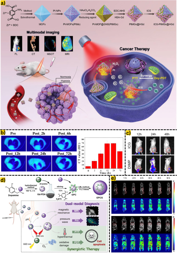
Metal-organic frameworks (MOFs), a burgeoning class of coordination polymers, have garnered significant attention due to their outstanding structure, porosity, and stability. They have been extensively studied in catalysis, energy storage, water harvesting, selective gas separation, and electrochemical applications. Recent advancements in post-synthetic strategies, surface functionality, and biocompatibility have expanded the application scope of MOFs, particularly in various biomedical fields. Herein, we review MOF-based nanomaterials bioimaging nanoplatforms in magnetic resonance imaging, computed tomography, and fluorescence imaging. MOFs serve as the foundation for biosensors, demonstrating efficiency in sensing H2O2, tumor biomarkers, microRNA, and living cancer cells. MOF-based carriers are well designed in drug delivery systems and anticancer treatment therapies. Additionally, we examine the challenges and prospects of MOFs in surface modification, release of metal ions, and interaction with intracellular components, as well as their toxicity and long-term effects.


Conflict of interest statement

There are no conflicts to declare.

Figures

Fig. 1
Fig. 1. A bibliometric analysis using VOSviewer software (a) and the number of publications reported on MOFs (b). Data retrieved from Scopus with keywords as “metal–organic framework” and “biomedical” between 2012 and 2022. Bibliometric pattern.
Fig. 2
Fig. 2. (a) Detailed synthesis of MOFs from Zr4+ and benzene-1,4-dicarboxylic acid by a solvothermal method and post-synthesis modification by loading indocyanine green. Reproduced from ref. with permission from Wiley-VCH Verlag, copyright 2020. (b) Tumor computed tomography images of mice after intravenous injection of the material at different time intervals (2, 6, 12, 24, and 72 h) are denoted by a green dashed ellipse. Computed tomography value (HU) of the material after each time interval. Reproduced from ref. with permission from Dove Medical Press Ltd, copyright 2020. (c) Fluorescence images of 4T1 tumor-bearing mice injected with indocyanine green and intracellular acidity-responsive polymeric MOF nanoparticles at different time points, tumors marked with red circles. Reproduced from ref. with permission from Elsevier, copyright 2021. (d) Phototherapy and synergistic treatment based on a single MOF material, Gd-MOF. Specifically, Gd from the MOF can support magnetic resonance imaging and Gd-polydopamine increases pressure waves for photothermal imaging application. In addition, loading chlorine6 onto the MOF surface also supports increased hyperthermal and oxidative damage to photothermal and photothermal therapies. Reproduced from ref. with permission from the Royal Society of Chemistry, copyright 2021. (e) Magnetic resonance images on the coronary plane of 4T1 mice in different stages after injection of MOF-based materials at tumor sites, liver and kidney. Tumors are marked with red circles. Reproduced from ref. with permission from the Royal Society of Chemistry, copyright 2021. Abbreviations: benzene-1,4-dicarboxylic acid, BDC; fluorescence imaging, FL; computed tomography, CT; multi-spectral optoacoustic tomography, MSOT; magnetic resonance imaging, MRI; intracellular acidity-responsive polymeric MOF nanoparticle, DIMP; photothermal therapy, PTT; photodynamic therapy, PDT; indocyanine green, ICG; photoacoustic imaging, PAI; photothermal imaging, PTI; multifunctional Gd-PDA-Ce6@Gd-MOF, GPCG; polydopamine, DPA.
Fig. 3
Fig. 3. (a) Principle of action of ICDSE for exosome enrichment from NSCLC patient plasma: Synthesis of engineered erythrocytes to obtain exosomes or exosomal cargo using CD63 aptamer biological affinity. (b) Exosomal miRNA installation based on the fabrication of a plasmonic biosensor based on supramolecular dendritic nanostructures and Zr MOF. Reproduced from ref. with permission from Elsevier, copyright 2023. (c) Colorimetric and electrochemical sensing method based on MOF-818 on a smartphone platform, developing a H2O2 sensing system from living cells. Reproduced from ref. with permission from Elsevier, copyright 2022.
Fig. 4
Fig. 4. (a) Manufacturing process of the MNPs/Zn-MOF modified electrode and H2O2 sensor from living cells released from drug stimulation and data transmission to the electrochemical station. Reproduced from ref. with permission from American Chemical Society, copyright 2022. (b) BUT-88-based DNA probe fabrication procedure for cytoplasmic miRNA-21 diagnosis in MCF-7 cells and simultaneous membrane-specific recognition of MUC-1. Reproduced from ref. with permission from Wiley-VCH Verlag, copyright 2020. (c) Schematic description of materials synthesis and use of Au@Cu-HHTP-NS modified electrodes to sense H2O2 in mitochondria from living human colon cells. Reproduced from ref. with permission from Elsevier, copyright 2022.
Fig. 5
Fig. 5. (a) During the synthesis process, a hollow porphyrinic ZIF-8-based composite is co-loaded with DOX and ICG and coated with cytomembrane for homotypic targeting and immune escape. As this material enters the bloodstream to the cancer cells under a pH response, they release ICGs and DOXs for PDT, PTT, and chemotherapy. Alternatively, the carrier can be irradiated with NIR to generate drugs and 1O2, which induce PDT effects and kills cancer cells. Reproduced from ref. with permission from American Chemical Society, copyright 2021. (b) A temozolomide drug delivery system based on MOF is injected directly into the mouse; the drug is transported through the bloodstream into nerve cells under the influence of ultrasound waves. Through this approach, the carrier easily penetrates the blood–brain barrier (BBB) and completely releases the drugs. Reproduced from ref. with permission from Dove Medical Press Ltd, copyright 2021.
Fig. 6
Fig. 6. (a) The mechanism of the MOF material for oral drug delivery, the drug passes through the stomach and is absorbed in the intestine. Reproduced from ref. with permission from Elsevier, copyright 2022. (b) The synthesis and functionalization of MOFs facilitate their absorption into the small intestinal cell wall, where they gradually undergo breakdown as they traverse each layer. Initially, they navigate through the upper tissue layer by shedding the PEG layer. Subsequently, they enter the lamina propria, a region supporting epithelial cells and facilitating the passage of blood vessels and nutrients. At this stage, the MOF material releases insulin drugs, which enter the bloodstream to perform their intended function. Reproduced from ref. with permission from American Chemical Society, copyright 2020. (c) The graph (left) shows the drug release mechanism from MOF materials in three types of media: deionized water, deionized water with 0.01 M H+ and deionized water with 0.01 M Na+. Accordingly, drug release was highest in deionized water with 0.01 M Na+, thus indicating that H+ could not trigger procainamide release from PEG@ZJU-64-NSN. This indicated that there was a strong interaction between the cationic procainamide and the anionic MOF framework. This strong interaction would be further enhanced by procainamide protonation under more acidic conditions. The graph (right) shows the drug release ability of the material in two simulated environments: the physiological environment and the acidic environment of the stomach. Reproduced from ref. with permission from Elsevier, copyright 2022.

References

    1. Shimizu K. Hibino S. Biros M. H. Irisawa T. Shimazu T. Int. J. Emerg. Med. 2021;14:2. doi: 10.1186/s12245-020-00316-7. - DOI - PMC - PubMed
    1. Tundisi L. L. Ataide J. A. Costa J. S. R. Coêlho D. de F. Liszbinski R. B. Lopes A. M. Oliveira-Nascimento L. de Jesus M. B. Jozala A. F. Ehrhardt C. Mazzola P. G. Colloids Surf., B. 2023;222:113043. doi: 10.1016/j.colsurfb.2022.113043. - DOI - PubMed
    1. Liu S. Tang J. Ji F. Lin W. Chen S. Gels. 2022;8:46. doi: 10.3390/gels8010046. - DOI - PMC - PubMed
    1. Rabeie B. Mahmoodi N. M. J. Colloid Interface Sci. 2024;654:495–522. doi: 10.1016/j.jcis.2023.10.060. - DOI - PubMed
    1. Ahmad N. Nordin N. A. H. M. Jaafar J. Ismail A. F. Ramli M. K. N. B. J. Environ. Chem. Eng. 2021;9:105887. doi: 10.1016/j.jece.2021.105887. - DOI