. 2023 Jun:11:109-156.
doi: 10.3114/fuse.2023.11.09. Epub 2023 Jun 16.

New and Interesting Fungi. 6

Affiliations

New and Interesting Fungi. 6

P W Crous et al. Fungal Syst Evol. 2023 Jun.

Abstract

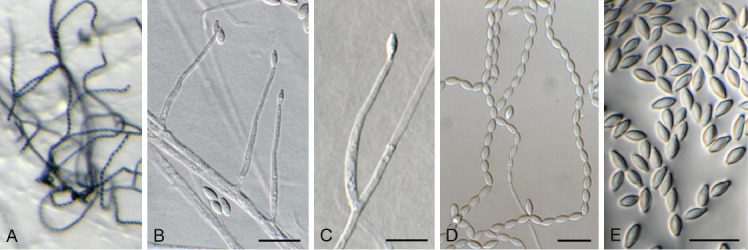
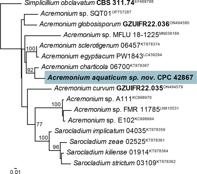
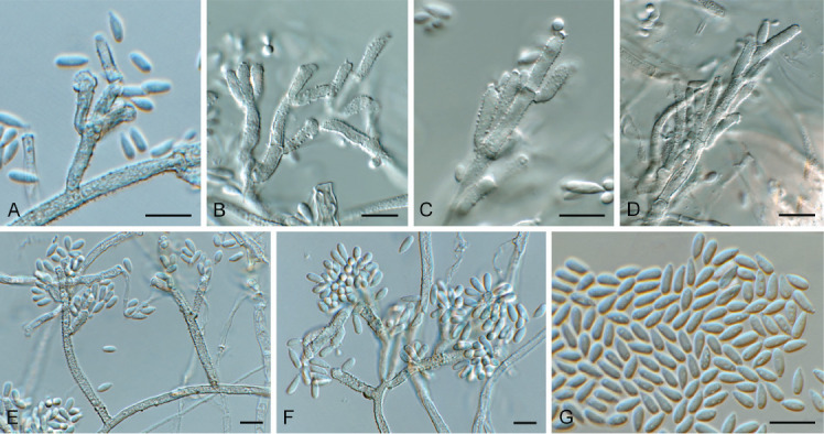
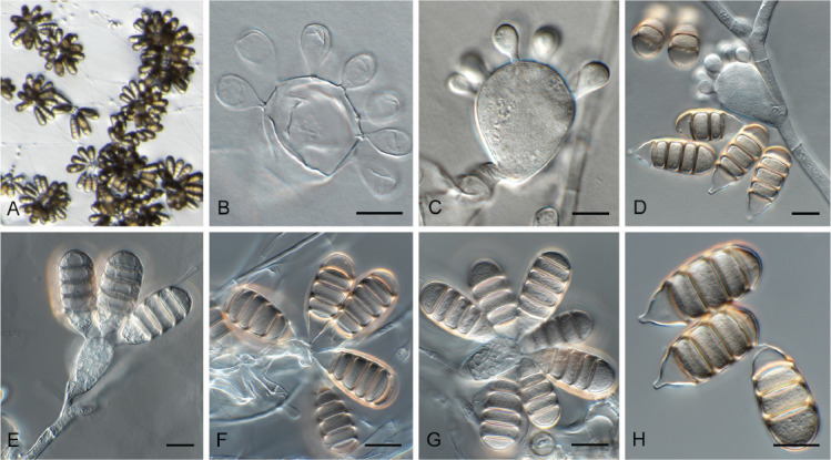
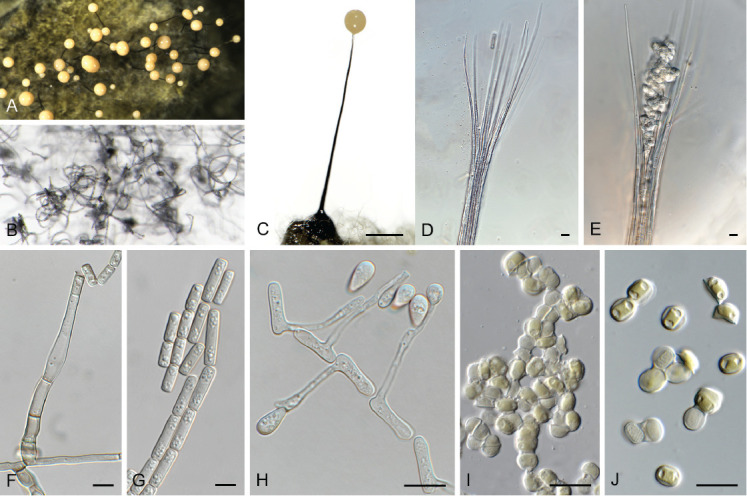
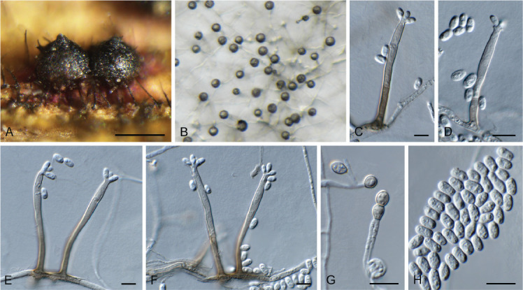
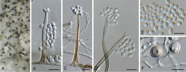
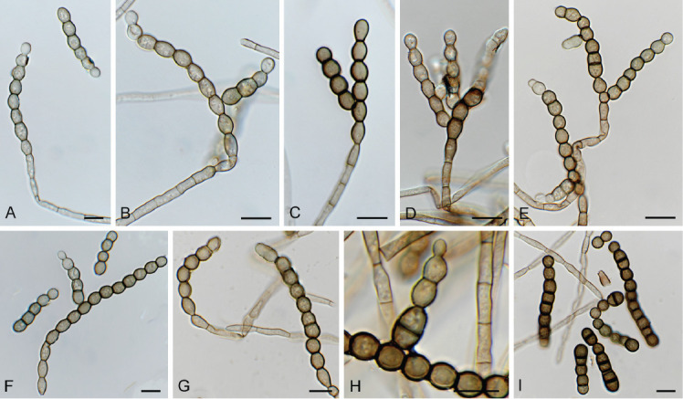
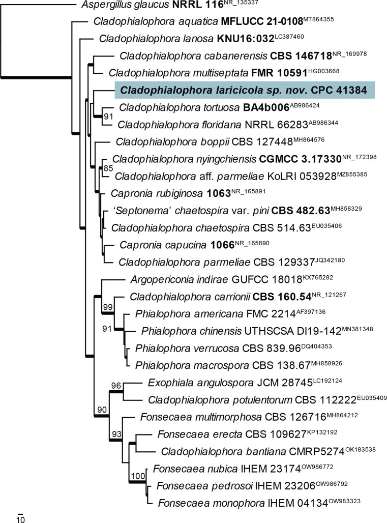
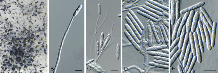
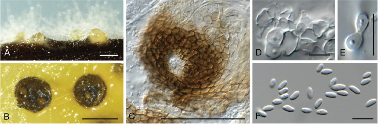
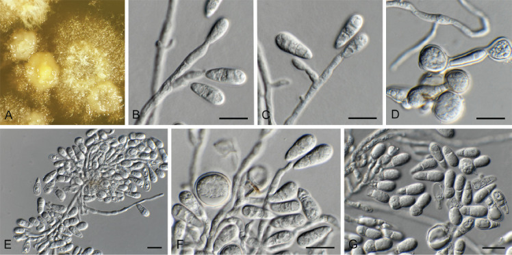
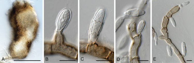
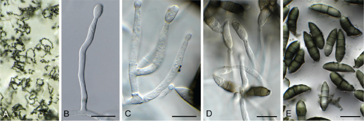
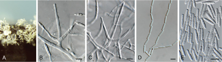
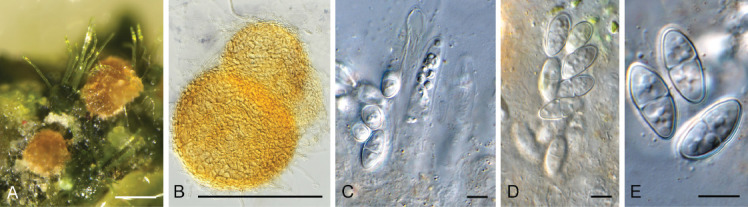
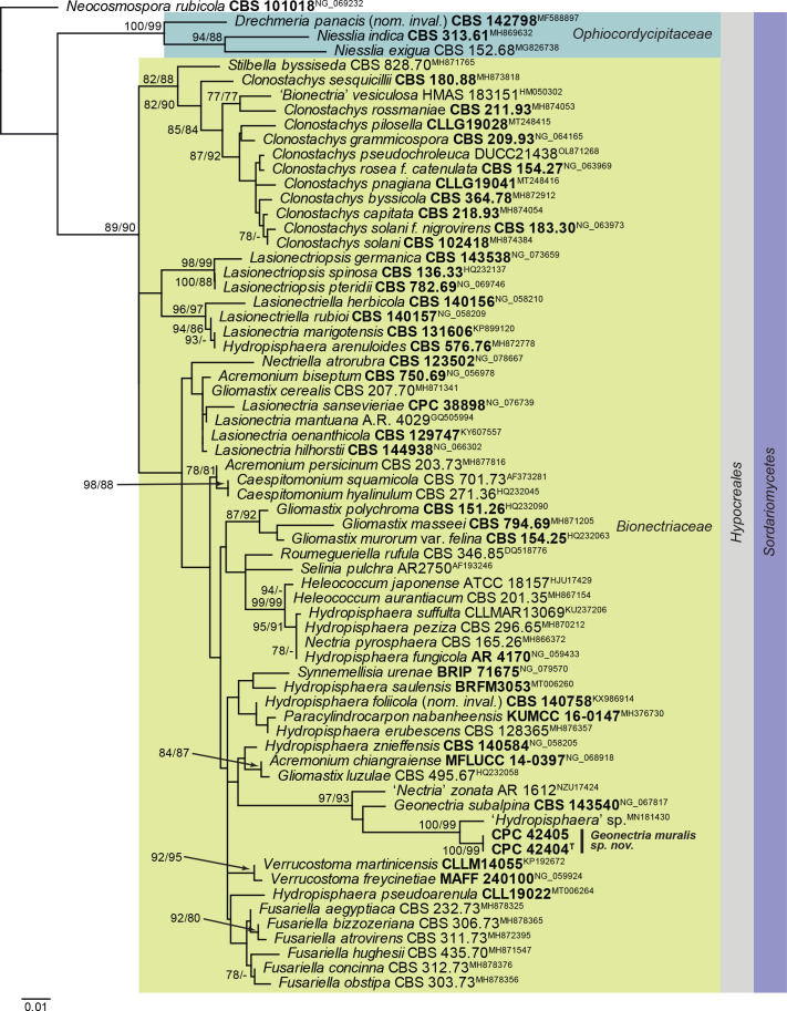
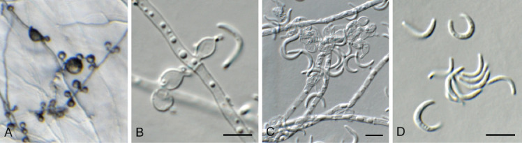
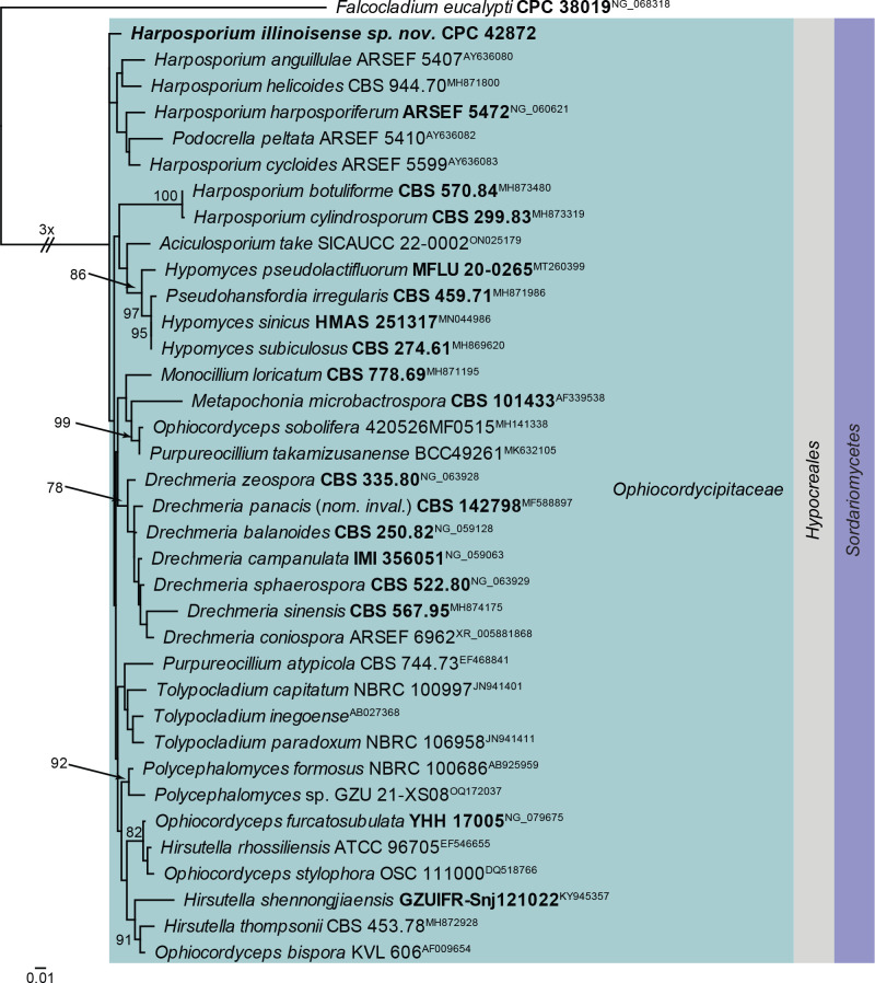
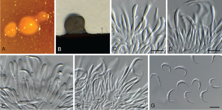
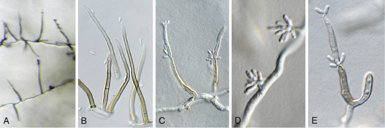
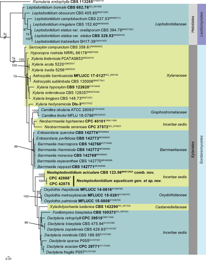
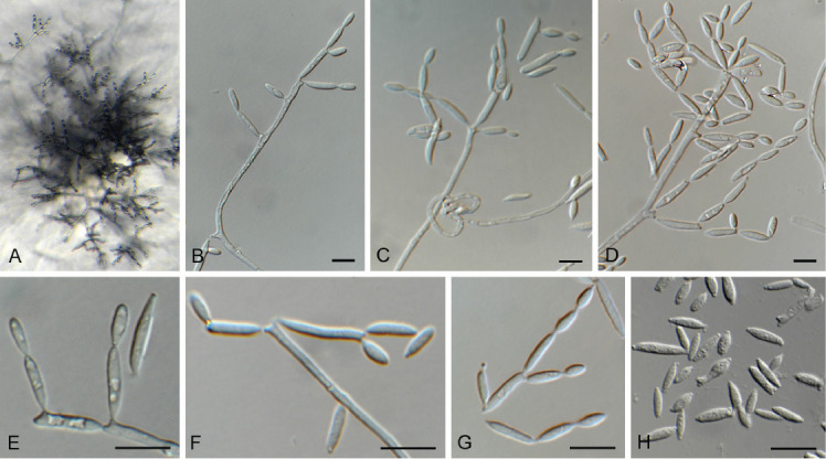
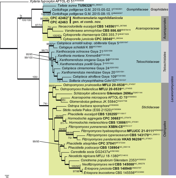
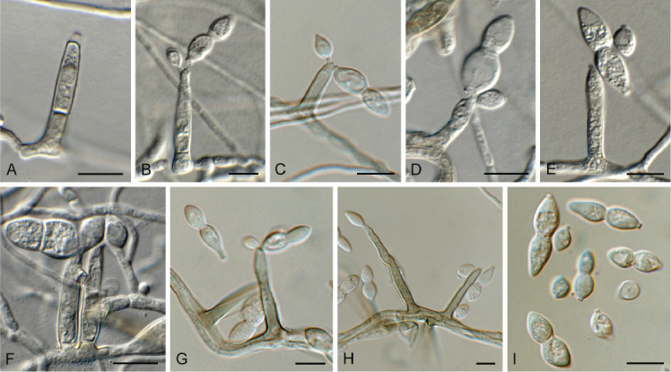
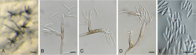
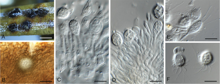
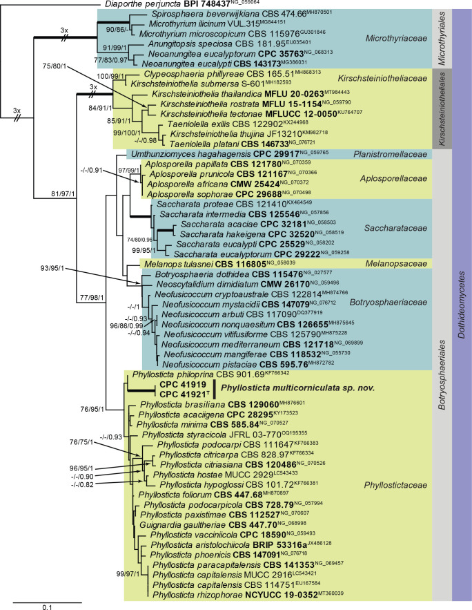
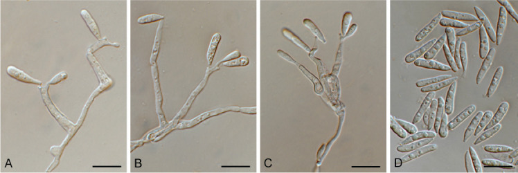
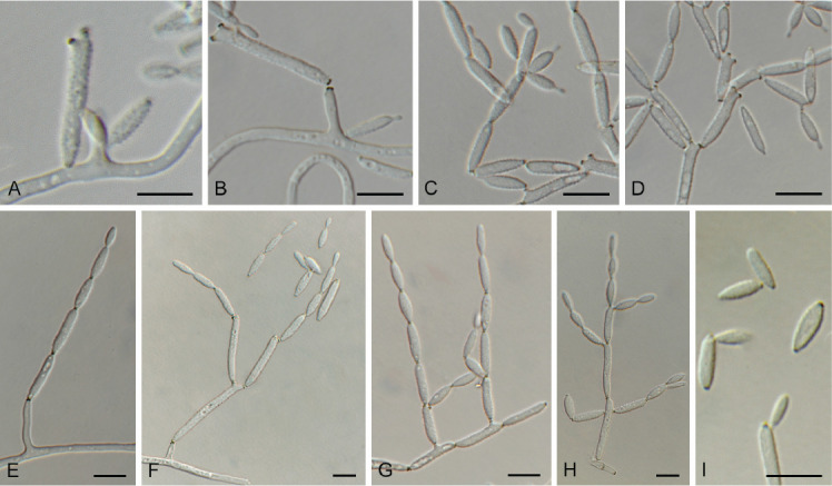
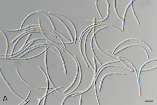
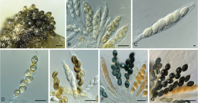
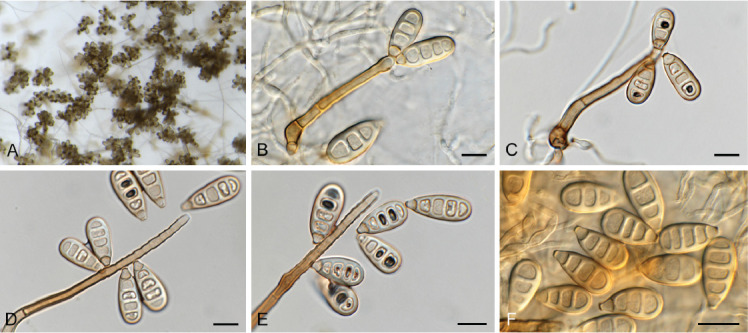
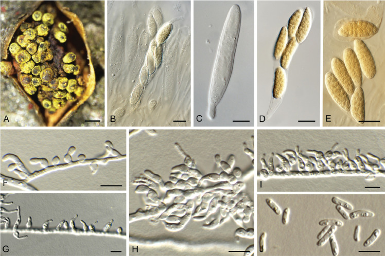
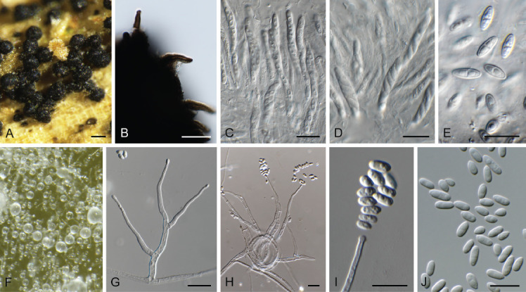
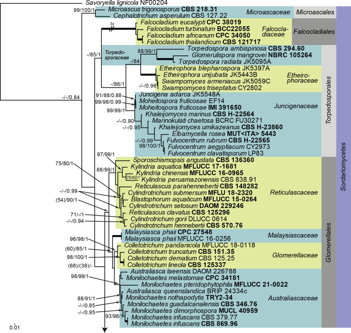
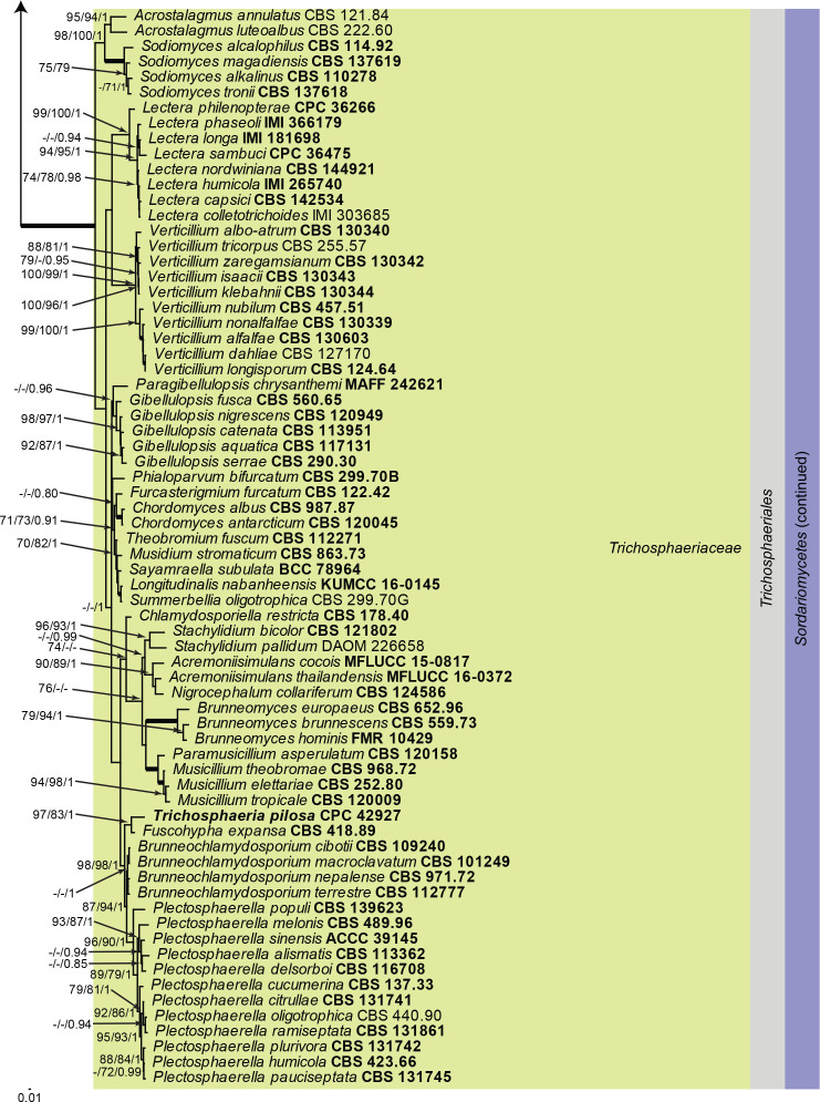
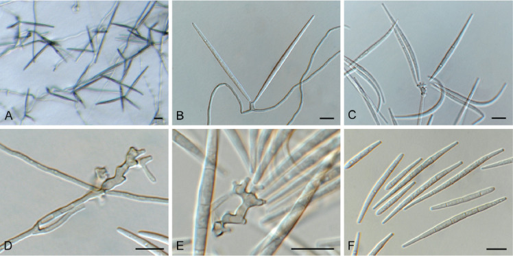
Three new genera, six new species, three combinations, six epitypes, and 25 interesting new host and / or geographical records are introduced in this study. New genera: Neoleptodontidium (based on Neoleptodontidium aquaticum), and Nothoramularia (based on Nothoramularia ragnhildianicola). New species: Acremonium aquaticum (from cooling pad water, USA, Cladophialophora laricicola (on dead wood of Larix sp., Netherlands), Cyphellophora neerlandica (on lichen on brick wall, Netherlands), Geonectria muralis (on moss growing on a wall, Netherlands), Harposporium illinoisense (from rockwool, USA), and Neoleptodontidium aquaticum (from hydroponic water, USA). New combinations: Cyphellophora deltoidea (based on Anthopsis deltoidea), Neoleptodontidium aciculare (based on Leptodontidium aciculare), and Nothoramularia ragnhildianicola (based on Ramularia ragnhildianicola). Epitypes: Cephaliophora tropica (from water, USA), Miricatena prunicola (on leaves of Prunus serotina, Netherlands), Nothoramularia ragnhildianicola (on Ragnhildiana ferruginea, parasitic on Artemisia vulgaris, Germany), Phyllosticta multicorniculata (on needles of Abietis balsamea, Canada), Thyronectria caraganae (on twigs of Caragana arborescens, Ukraine), and Trichosphaeria pilosa (on decayed Salix branch, Netherlands). Furthermore, the higher order phylogeny of three genera regarded as incertae sedis is resolved, namely Cephaliophora (Ascodesmidaceae, Pezizales), Miricatena (Helotiales, Leotiomycetes), and Trichosphaeria (Trichosphaeriaceae, Trichosphaeriales), with Trichosphaeriaceae being an older name for Plectosphaerellaceae. Citation: Crous PW, Akulov A, Balashov S, Boers J, Braun U, Castillo J, Delgado MA, Denman S, Erhard A, Gusella G, Jurjević Ž, Kruse J, Malloch DW, Osieck ER, Polizzi G, Schumacher RK, Slootweg E, Starink-Willemse M, van Iperen AL, Verkley GJM, Groenewald JZ (2023). New and Interesting Fungi. 6. Fungal Systematics and Evolution 11: 109-156. doi: 10.3114/fuse.2023.11.09.

Keywords: ITS barcodes; biodiversity; multi-gene phylogeny; new taxa; systematics; typification.


Conflict of interest statement

Conflict of interest: The authors declare that there is no conflict of interest.

Figures

Fig. 1.
Fig. 1.
Acremonium aquaticum (CPC 42867). A. Colony on SNA. B–D. Conidiogenous cells giving rise to chains of conidia. E. Conidia. Scale bars = 10 μm.
Fig. 2.
Fig. 2.
Consensus phylogram (50 % majority rule) obtained from the maximum likelihood analysis with IQ-TREE v. 2.1.3 of the Acremonium tef1 (second part) nucleotide alignment. Bootstrap support values (> 75 %) from 1 000 non-parametric bootstrap replicates are shown at the nodes. Culture collection or voucher numbers and GenBank accession numbers (superscript) are indicated for all species. Sequences derived from material with a type status are indicated with a culture or voucher number highlighted with bold face. The tree was rooted to Simplicillium obclavatum (culture CBS 311.74; GenBank EF468798) and the species treated here is highlighted with bold face. The scale bar indicates the expected number of changes per site.
Fig. 3.
Fig. 3.
Acrostalagmus luteoalbus (CPC 43187). A–G. Conidiophores with conidiogenous cells giving rise to conidia. H. Conidia. Scale bars = 10 μm.
Fig. 4.
Fig. 4.
Appendopyricularia juncicola (CPC 44053). A–D. Conidiophores with conidiogenous cells giving rise to conidia. E. Conidia. Scale bars = 10 μm.
Fig. 5.
Fig. 5.
Biscogniauxia anceps (CPC 43197). A–F. Conidiophores with conidiogenous cells giving rise to conidia. G. Conidia. Scale bars = 10 μm.
Fig. 6.
Fig. 6.
Cephaliophora tropica (CPC 42877). A–G. Conidiophores with conidiogenous cells giving rise to conidia. H. Conidia. Scale bars = 10 μm.
Fig. 7.
Fig. 7.
Ceratocystis ficicola (CPC 44213). A. Colony on PDA. B. Colony on SNA. C. Ascoma exuding ascospores. D, E. Ostiolar hyphae. F, G. Subcylindrical endoconidia. H. Aleuroconidia. I, J. Ascospores. Scale bars: C = 300 μm, all others = 10 μm.
Fig. 8.
Fig. 8.
Chloridium caudigerum (CPC 42899). A. Ascomata. B–F. Conidiophores with conidiogenous cells giving rise to conidia. G. Chlamydospores. H. Conidia. Scale bars: A = 300 μm, all others = 10 μm.
Fig. 9.
Fig. 9.
Chloridium gamsii (CPC 41933). A–D. Conidiophores with conidiogenous cells giving rise to conidia. E. Conidia. F. Chlamydospores. Scale bars = 10 μm.
Fig. 10.
Fig. 10.
Cladophialophora laricicola (CPC 41384). A–E, G. Conidiophores with conidiogenous cells giving rise to conidia. F, H, I. Conidia. Scale bars = 10 μm.
Fig. 11.
Fig. 11.
The first of four equally most parsimonious trees obtained from a ‘Cladophialophora’ ITS sequence alignment. The scale bar indicates the number of changes and the numbers at the nodes represent bootstrap support values (>74 %) based on 1 000 resamplings. Branches that appear in the strict consensus tree are highlighted by thickened lines. Culture collection or voucher numbers and GenBank accession numbers (superscript) are indicated for all species. Sequences derived from material with a type status are indicated with a culture or voucher number highlighted with bold face. The tree was rooted to Aspergillus glaucus (culture NRRL 116; GenBank NR_135337) and the species treated here is highlighted with bold face.
Fig. 12.
Fig. 12.
Cylindromonium eugeniicola (CPC 43326). A. Colony on SNA. B–D. Conidiophores with conidiogenous cells giving rise to conidia. E. Conidia. Scale bars = 10 μm.
Fig. 13.
Fig. 13.
Cyphellophora neerlandica (CPC 42634). A, B. Conidiogenous cells giving rise to conidia. C. Conidia. Scale bars = 10 μm.
Fig. 14.
Fig. 14.
Consensus phylogram (50 % majority rule) obtained from the maximum likelihood analysis with IQ-TREE v. 2.1.3 of the Anthopsis / Cyphellophora ITS nucleotide alignment. Maximum likelihood (> 74 %) and maximum parsimony (> 74 %) bootstrap support values from 1 000 non-parametric bootstrap replicates are shown at the nodes. Culture collection or voucher numbers and GenBank accession numbers (superscript) are indicated for all species. Sequences derived from material with a type status are indicated with a culture or voucher number highlighted with bold face. The tree was rooted to Capronia rubiginosa (culture BBB 536; GenBank NR_165891) and the species treated here is highlighted with bold face. The scale bar indicates the expected number of changes per site.
Fig. 15.
Fig. 15.
Didymella brevipilosa (CPC 41600). A. Conidiomata on SNA. B. Conidiomata on OA. C. Conidioma with ostiole. D, E. Conidiogenous cells. F. Conidia. Scale bars = 10 μm.
Fig. 16.
Fig. 16.
Drepanopeziza populi-albae (CPC 42336). A. Colony on MEA. B–G. Conidiophores with conidiogenous cells giving rise to conidia (note germinating conidia in D). Scale bars = 10 μm.
Fig. 17.
Fig. 17.
Endoconidioma populi (CPC 41602). A. Sclerotium-like body on SNA. B–E. Conidiogenous cells giving rise to conidia. Scale bars = 10 μm.
Fig. 18.
Fig. 18.
Fusariella atrovirens (CPC 43304). A. Colony on OA. B–D. Conidiogenous cells giving rise to conidia. E. Conidia. Scale bars = 10 μm.
Fig. 19.
Fig. 19.
Fusariella hughesii (CPC 41594). A. Sporulation on PNA. B, C. Conidiophores and conidiogenous cells giving rise to conidia. D, E. Conidia. Scale bars = 10 μm.
Fig. 20.
Fig. 20.
Geonectria muralis (CPC 42404). A. Ascomata in vivo. B. Ascomata in vitro. C, D. Asci with ascospores. E. Ascospores. Scale bars: A, B = 180 μm, all others = 10 μm.
Fig. 21.
Fig. 21.
Consensus phylogram (50 % majority rule) obtained from the maximum likelihood analysis with IQ-TREE v. 2.1.3 of the Hypocreales LSU nucleotide alignment. Maximum likelihood (> 74 %) and maximum parsimony (> 74 %) bootstrap support values from 1 000 non-parametric bootstrap replicates are shown at the nodes. Culture collection or voucher numbers and GenBank accession numbers (superscript) are indicated for all species. Sequences derived from material with a type status are indicated with a culture or voucher number highlighted with bold face. The tree was rooted to Neocosmospora rubicola (culture CBS 101018; GenBank NG_069232) and the species treated here is highlighted with bold face. The families, order and class are shown in coloured blocks to the right of the tree. The scale bar indicates the expected number of changes per site.
Fig. 22.
Fig. 22.
Harposporium illinoisense (CPC 42872). A. Colony on SNA. B, C. Conidiogenous cells giving rise to conidia. D. Conidia. Scale bars = 10 μm.
Fig. 23.
Fig. 23.
Consensus phylogram (50 % majority rule) obtained from the maximum likelihood analysis with IQ-TREE v. 2.1.3 of the Ophiocordycipitaceae LSU nucleotide alignment. Bootstrap support values (> 75 %) from 1 000 non-parametric bootstrap replicates are shown at the nodes. Culture collection or voucher numbers and GenBank accession numbers (superscript) are indicated for all species. Sequences derived from material with a type status are indicated with a culture or voucher number highlighted with bold face. The tree was rooted to Falcocladium eucalypti (culture CPC 38019; GenBank NG_068318) and the species treated here is highlighted with bold face. The family, order and class are shown in coloured blocks to the right of the tree. The scale bar indicates the expected number of changes per site.
Fig. 24.
Fig. 24.
Microcera physciae (CPC 42638). A. Colony sporulating on OA. B. Colony sporulating on PNA. C–F. Conidiophores and conidiogenous cells giving rise to conidia. G. Conidia. Scale bars = 10 μm.
Fig. 25.
Fig. 25.
Miricatena prunicola (CPC 42627). A, B. Leaf spots. C. Conidiogenous cells giving rise to secondary conidia. D. Secondary conidia. E, F. Primary conidia. G, H. Primary conidia giving rise to secondary conidia. Scale bars -= 10 μm.
Fig. 26.
Fig. 26.
Neoleptodontidium aquaticum (CPC 42868). A. Colony on SNA. B–E. Conidiophores and conidiogenous cells giving rise to conidia. Scale bars = 10 μm.
Fig. 27.
Fig. 27.
Consensus phylogram (50 % majority rule) obtained from the maximum likelihood analysis with IQ-TREE v. 2.1.3 of the Xylariales LSU nucleotide alignment. Maximum likelihood (> 74 %) bootstrap support values from 1 000 non-parametric bootstrap replicates are shown at the nodes. Culture collection or voucher numbers and GenBank accession numbers (superscript) are indicated for all species. Sequences derived from material with a type status are indicated with a culture or voucher number highlighted with bold face. The tree was rooted to Ramularia endophylla (culture CBS 113265; GenBank AY490776) and the species treated here are highlighted with bold face. The families, orders and classes are shown in coloured blocks to the right of the tree. The scale bar indicates the expected number of changes per site.
Fig. 28.
Fig. 28.
Nothoramularia ragnhildianicola (CPC 42462). A. Colony on SNA. B–D, F. Conidiophores and conidiogenous cells giving rise to conidia. E, G, H. Conidia. Scale bars = 10 μm.
Fig. 29.
Fig. 29.
The first of two equally most parsimonious trees obtained from the Lecanoromycetes LSU sequence alignment. The scale bar indicates the number of changes and the numbers at the nodes represent maximum parsimony (>70 %) and maximum likelihood (> 70 %) bootstrap support values from 1 000 non-parametric bootstrap replicates. Branches that appear in the strict consensus tree are highlighted by thickened lines. Culture collection or voucher numbers and GenBank accession numbers (superscript) are indicated for all species. Sequences derived from material with a type status are indicated with a culture or voucher number highlighted with bold face. The tree was rooted to Xylaria hypoxylon (AFTOL-ID 51; GenBank AY544648) and the species treated here is highlighted with bold face. The families, orders and class are shown in coloured blocks to the right of the tree.
Fig. 30.
Fig. 30.
Paradissoconium narthecii (CPC 42494). A. Conidiophore. B–H. Conidiophores and conidiogenous cells giving rise to primary and secondary conidia. I. One-septate primary and aseptate secondary conidia. Scale bars = 10 μm.
Fig. 31.
Fig. 31.
Phomatospora endopteris (CPC 41832). A. Colony on SNA. B–D. Conidiophores and conidiogenous cells giving rise to conidia. E. Conidia. Scale bars = 10 μm.
Fig. 32.
Fig. 32.
Phyllosticta multicorniculata (CPC 41921). A. Erumpent conidiomata on Abies needle. B. Conidioma with central ostiole. C, D. Conidiogenous cells giving rise to conidia. E, F. Conidia with multiple apical appendages. Scale bars: A = 200 μm, all others = 10 μm.
Fig. 33.
Fig. 33.
Consensus phylogram (50 % majority rule) obtained from the maximum likelihood analysis with IQ-TREE v. 2.1.3 of the Phyllostictaceae and related families LSU nucleotide alignment. Maximum likelihood (> 74 %) and maximum parsimony (> 74 %) bootstrap support values from 1 000 non-parametric bootstrap replicates, and Bayesian posterior probabilities (> 0.84), are shown at the nodes. Thickened lines represent nodes which received full support (100 % / 100 % / 1) from all three analyses. Culture collection or voucher numbers and GenBank accession numbers (superscript) are indicated for all species. Sequences derived from material with a type status are indicated with a culture or voucher number highlighted with bold face. The tree was rooted to Diaporthe perjuncta (voucher BPI 748437; GenBank NG_059064) and the species treated here is highlighted with bold face. The families, orders and class are shown in coloured blocks to the right of the tree. The scale bar indicates the expected number of changes per site.
Fig. 34.
Fig. 34.
Pleurotheciella aquatica (CPC 44105). A–C. Conidiophores and conidiogenous cells giving rise to conidia. D. Conidia. Scale bars = 10 μm.
Fig. 35.
Fig. 35.
Ramularia pistaciae (CPC 44067). A–H. Conidiophores and conidiogenous cells giving rise to branched conidial chains. I. Conidia. Scale bars = 10 μm.
Fig. 36.
Fig. 36.
Ruptoseptoria unedonis (CPC 44069). Conidia. Scale bar = 10 μm.
Fig. 37.
Fig. 37.
Schizothecium conica (CPC 44110). A. Ascomata on SNA. B–G. Asci with ascospores. Scale bars: A = 180 μm, all others = 10 μm.
Fig. 38.
Fig. 38.
Sporidesmiella pini (CPC 41495). A. Colony on SNA. B–E. Conidiophores and conidiogenous cells giving rise to conidia. F. Conidia. Scale bars = 10 μm.
Fig. 39.
Fig. 39.
Thyronectria caraganae (CPC 41504). A. Ascomata erumpent from host tissue. B, C. Asci. D, E. Ascospores. F–I. Conidiophores and conidiogenous cells giving rise to conidia. J. Conidia. Scale bars: A = 180 μm, all others = 10 μm.
Fig. 40.
Fig. 40.
Trichosphaeria pilosa (CPC 42927). A. Ascomata on host tissue. B. Ascomatal wall with setae. C, D. Asci. E. Ascospores. F. Colony on OA. G–I. Conidiophores and conidiogenous cells giving rise to conidia. J. Conidia. Scale bars: A = 180 μm, all others = 10 μm.
Fig. 41.
Fig. 41.
Consensus phylogram (50 % majority rule) obtained from the maximum likelihood analysis with IQ-TREE v. 2.1.3 of the Sordariomycetes ITS/LSU/tef1 nucleotide alignment. Maximum likelihood (> 74 %) and RAxML (> 74 %) bootstrap support values from 1 000 non-parametric bootstrap replicates, and Bayesian posterior probabilities (> 0.84), are shown at the nodes. Thickened lines represent nodes which received full support (100 % / 100 % / 1) from all three analyses. Culture collection or voucher numbers are indicated for all species. Sequences derived from material with a type status are indicated with a culture or voucher number highlighted with bold face. GenBank accession numbers of the sequences used in the alignment are listed in supplementary Table S2. The tree was rooted to Savoryella lignicola (strain NF00204) and the species treated here is highlighted with bold face. The families, orders and class are shown in coloured blocks to the right of the tree. The scale bar indicates the expected number of changes per site.
Fig. 41.
Fig. 41.
Consensus phylogram (50 % majority rule) obtained from the maximum likelihood analysis with IQ-TREE v. 2.1.3 of the Sordariomycetes ITS/LSU/tef1 nucleotide alignment. Maximum likelihood (> 74 %) and RAxML (> 74 %) bootstrap support values from 1 000 non-parametric bootstrap replicates, and Bayesian posterior probabilities (> 0.84), are shown at the nodes. Thickened lines represent nodes which received full support (100 % / 100 % / 1) from all three analyses. Culture collection or voucher numbers are indicated for all species. Sequences derived from material with a type status are indicated with a culture or voucher number highlighted with bold face. GenBank accession numbers of the sequences used in the alignment are listed in supplementary Table S2. The tree was rooted to Savoryella lignicola (strain NF00204) and the species treated here is highlighted with bold face. The families, orders and class are shown in coloured blocks to the right of the tree. The scale bar indicates the expected number of changes per site.
Fig. 42.
Fig. 42.
Zaanenomyces versatilis (CPC 42831). A. Colony on SNA. B–E. Conidiophores and conidiogenous cells giving rise to conidia. F. Conidia. Scale bars = 10 μm.

References

    1. Andersson MA, Salo J, Mikkola R, et al. (2021). Melinacidin-producing Acrostalagmus luteoalbus, a major constituent of mixed mycobiota contaminating insulation material in an outdoor wall. Pathogens 10: 843. - PMC - PubMed
    1. Bahmani Z, Abdollahzadeh J, Amini J. et al. (2021). Biscogniauxia rosacearum the charcoal canker agent as a pathogen associated with grapevine trunk diseases in Zagros region of Iran. Scientific Reports 11: 14098. - PMC - PubMed
    1. Barr ME. (1990). Prodromus to nonlichenized, pyrenomycetous members of class Hymenoascomycetes. Mycotaxon 39: 43–184.
    1. Barr ME, Cannon P. (1994). Discussion 3: Calosphaeriales, Clavicipitales, Coryneliales, Diaporthales, Diatrypales, Halosphaeriales, Hypocreales, Meliolales, Ophiostomatales, Phyllachorales, Sordariales, Trichosphaeriales, and Xylariales. Ascomycete Systematics Problems and Perspectives in the Nineties (Hawksworth DL, ed.) New York & London: Plenum Press: 371–378.
    1. Bell A, Mahoney DP. (1995). Coprophilous fungi from New Zealand. I. Podospora species with swollen agglutinated perithecial hairs. Mycologia 87: 375–396.

LinkOut - more resources