. 2024 Apr;13(4):e12421.
doi: 10.1002/jev2.12421.

Identification and validation of extracellular vesicle reference genes for the normalization of RT-qPCR data

Affiliations

Identification and validation of extracellular vesicle reference genes for the normalization of RT-qPCR data

Cláudio Pinheiro et al. J Extracell Vesicles. 2024 Apr.

Abstract

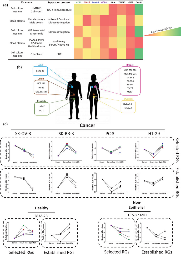
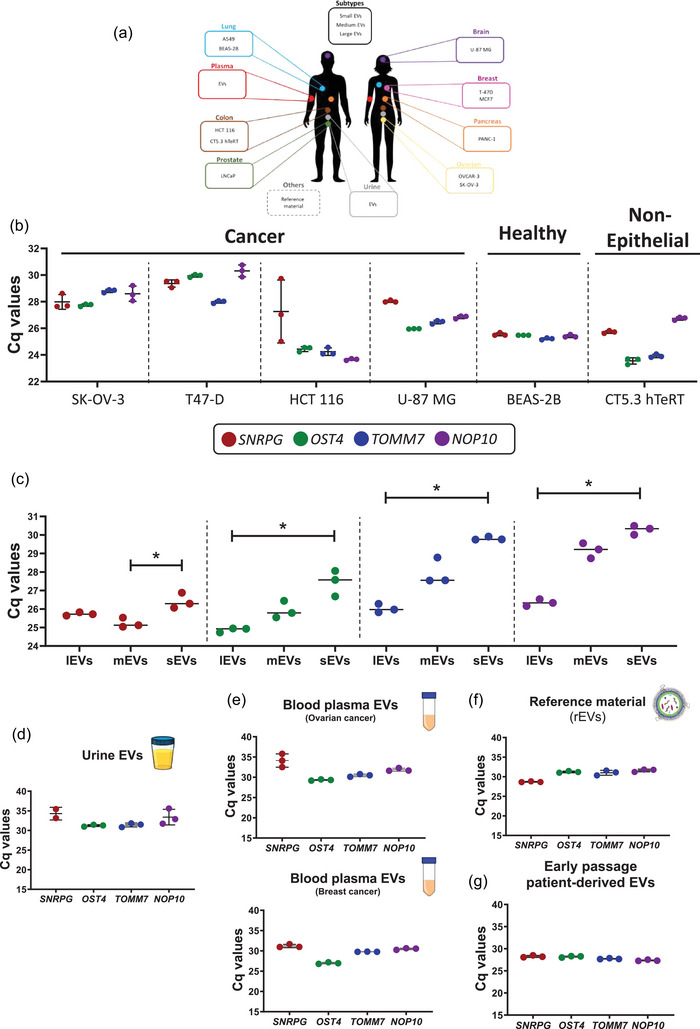
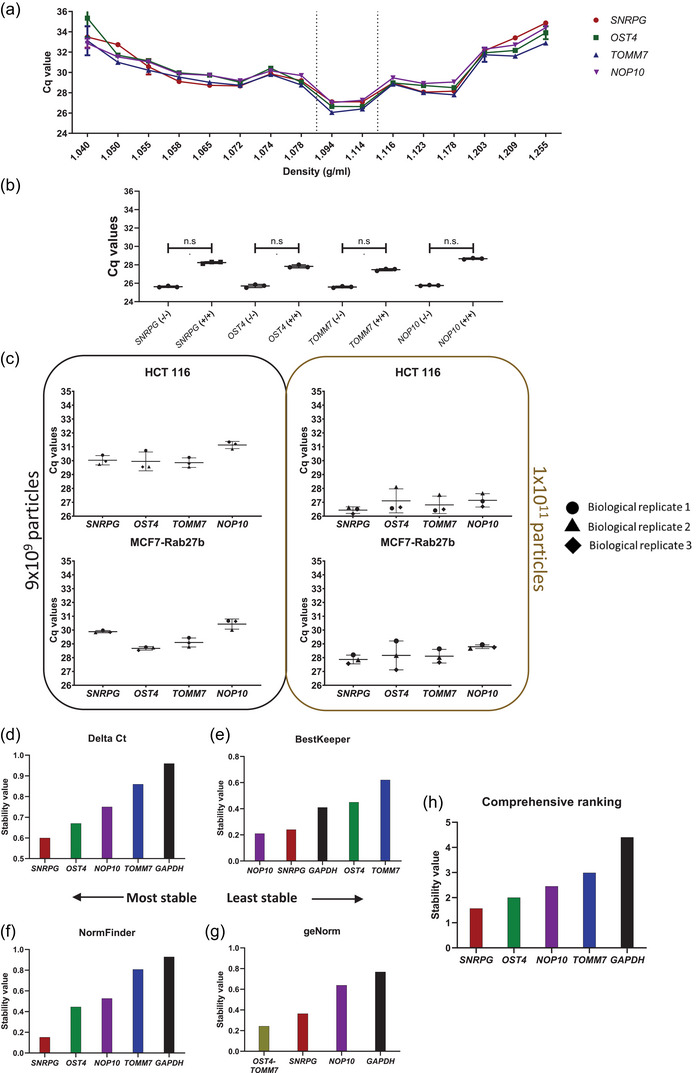
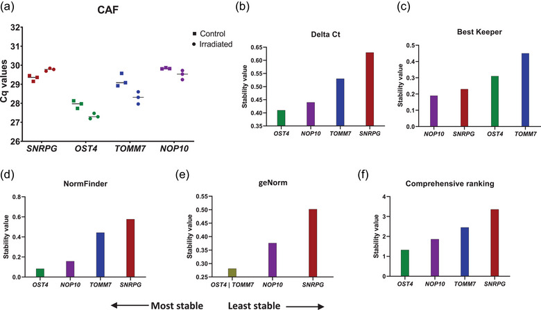
Extracellular vesicles (EVs) contain a plethora of biomolecules, including nucleic acids, with diverse diagnostic and therapeutic application potential. Although reverse transcription-quantitative PCR (RT-qPCR) is the most widely applied laboratory technique to evaluate gene expression, its applicability in EV research is challenged by the lack of universal and stably present reference genes (RGs). In this study, we identify, validate and establish SNRPG, OST4, TOMM7 and NOP10 as RGs for the normalization of EV-associated genes by RT-qPCR. We show the stable presence of SNRPG, OST4, TOMM7 and NOP10 in multiple cell lines and their secreted EVs (n = 12) under different (patho)physiological conditions as well as in human-derived biofluids (n = 3). Enzymatic treatments confirm the presence of SNRPG, OST4, TOMM7 and NOP10 inside EVs. In addition, the four EV-associated RGs are stably detected in a size-range of EV subpopulations. RefFinder analysis reveals that SNRPG, OST4, TOMM7 and NOP10 are more stable compared to RGs established specifically for cultured cells or tissues such as HMBS, YWHAZ, SDHA and GAPDH. In summary, we present four universal and stably present EV-associated RGs to enable normalization and thus steer the implementation of RT-qPCR for the analysis of EV-associated RNA cargo for research or clinical applications.

Keywords: RNase; blood; conditioned medium; exosomes; extracellular vesicles; mRNA; microvesicles; protease; reference genes; tissue; urine.


Conflict of interest statement

A.H., O.D.W., J.V. and P.M. are inventors on the patent application covering the rEV technology (WO2019091964).

Figures

FIGURE 1
FIGURE 1
Identification of SNRPG, OST4, TOMM7, NOP10 and their evaluation versus established cell line and tissue reference genes (RGs) in cell lines of diverse origin. (a) Evaluation of the presence of identified (EV) and established (cell lines and tissue) RGs in five publicly available EV RNA datasets. Heatmap is based on relative abundance within each respective study. (b) Selected panel of 15 cell lines for the evaluation of RGs. (c) Expression profiles obtained by plotting the Cq value deviations relative to the serum condition (ΔCq serum free = avg Cq (RG) in serum – avg Cq (gene) in serum free; ΔCq opti‐MEM = avg Cq (gene) in serum free – avg Cq (gene) in opti‐MEM; data was normalized to average Cq value per RG in serum). Cell lines were cultured for 24 h under three different conditions: 10% serum (circles); serum‐ free (squares) and in Opti‐MEM medium (inverted triangle). Results are represented as expression profiles of tested genes in average delta Cq values after analysis of three technical replicates. CP, chronic pancreatitis; PDAC, pancreatic ductal adenocarcinoma.
FIGURE 2
FIGURE 2
Identification of SNRPG, OST4, TOMM7 and NOP10 in EV preparations from diverse origins. (a) Selected panel of different origins from which EVs were prepared. (b) RT‐qPCR analysis of EVs prepared from media conditioned by cancer, healthy or non‐epithelial cell lines using DGUC. (c) RT‐qPCR analysis of a size range of EV subpopulations fractionated media conditioned by MCF7 cells using dual SEC (Kruskal–Wallis test, adjusted p‐value for Dunns's multiple comparison test = 0.0219). (d–f) RT‐qPCR analysis of EVs prepared from urine by DGUC (pool of healthy donors (n = 3)) (d), (e) EVs prepared from blood plasma using SEC (ovarian (n = 2) and breast (n = 2) cancer patients), (f) EV reference material (rEVs) and (g) early passage patient‐derived sarcoma cells. Results were obtained by analysis of three technical replicates. * Adjusted p‐value >0.05.
FIGURE 3
FIGURE 3
Validation of SNRPG, OST4, TOMM7 and NOP10 as suitable EV reference genes (RGs) using enzymatic treatments, biological replicates and stability algorithms. (a) RT‐qPCR analysis of all density fractions prepared from media conditioned by HCT 116 cells using DGUC. (b) RT‐qPCR analysis of SNRPG, OST4, TOMM7 and NOP10 in all density fractions prepared from media conditioned by HCT 116 cells using DGUC prior (blue) and after (red) proteinase K and RNase A treatment. Dotted lines represent average Cq values of selected RGs in soluble protein fractions (1.06–1.07 g/mL). (c) Intrinsic biological variability of EVs prepared from media conditioned by HCT 116 or MCF7 cells using DGUC in three biological replicates. Normalization to particle number was performed via nanoparticle tracking analysis (NTA). Biological results are shown as average Cq values of three technical replicates. (d–h) Stability evaluation using the RefFinder tool of EV RGs versus the established tissue and cell lines RG GAPDH in EVs prepared from media conditioned by LnCap via DGUC. Results of four algorithms are shown, (d) Delta Ct, (e) BestKeeper, (f) NormFinder and (g) geNorm alongside their respective (h) geometric mean.
FIGURE 4
FIGURE 4
Validation of SNRPG, OST4, TOMM7 and NOP10 as suitable reference genes using destabilizing conditions. (a) RT‐qPCR analysis of EVs prepared from media conditioned by CAFs (irradiated (10 Gy) or not) by DGUC in three technical replicates. Stability analysis of SNRPG, OST4, TOMM7 and NOP10 was performed using the RefFinder tool. Results of four algorithms are shown, (b) Delta Ct, (c) BestKeeper, (d) NormFinder and (e) geNorm alongside their respective (f) geometric mean.

References

    1. Abumaghaid, M. M. , Abdelazim, A. M. , Belali, T. M. , Alhujaily, M. , & Saadeldin, I. M. (2022). Shuttle transfer of mRNA transcripts via extracellular vesicles from male reproductive tract cells to the cumulus‐oocyte complex in rabbits (Oryctolagus cuniculus). Frontiers in Veterinary Science, 9, 816080. - PMC - PubMed
    1. Andersen, C. L. , Jensen, J. L. , & Ørntoft, T. F. (2004). Normalization of real‐time quantitative reverse transcription‐PCR data: A model‐based variance estimation approach to identify genes suited for normalization, applied to bladder and colon cancer data sets. Cancer Research, 64, 5245–5250. - PubMed
    1. Batagov, A. O. , & Kurochkin, I. V. (2013). Exosomes secreted by human cells transport largely mRNA fragments that are enriched in the 3'‐untranslated regions. Biology Direct, 8, 12. - PMC - PubMed
    1. Battich, N. , Stoeger, T. , & Pelkmans, L. (2015). Control of transcript variability in single mammalian cells. Cell, 163, 1596–1610. - PubMed
    1. Bustin, S. A. , Benes, V. , Garson, J. A. , Hellemans, J. , Huggett, J. , Kubista, M. , Mueller, R. , Nolan, T. , Pfaffl, M. W. , Shipley, G. L. , Vandesompele, J. , & Wittwer, C. T. (2009). The MIQE guidelines: Minimum information for publication of quantitative real‐time PCR experiments. Clinical Chemistry, 55, 611–622. - PubMed

LinkOut - more resources