. 2024 Mar;105(3):001970.
doi: 10.1099/jgv.0.001970.

Acute Rift Valley fever virus infection induces inflammatory cytokines and cell death in ex vivo rat brain slice culture

Affiliations

Acute Rift Valley fever virus infection induces inflammatory cytokines and cell death in ex vivo rat brain slice culture

Kaleigh A Connors et al. J Gen Virol. 2024 Mar.

Abstract

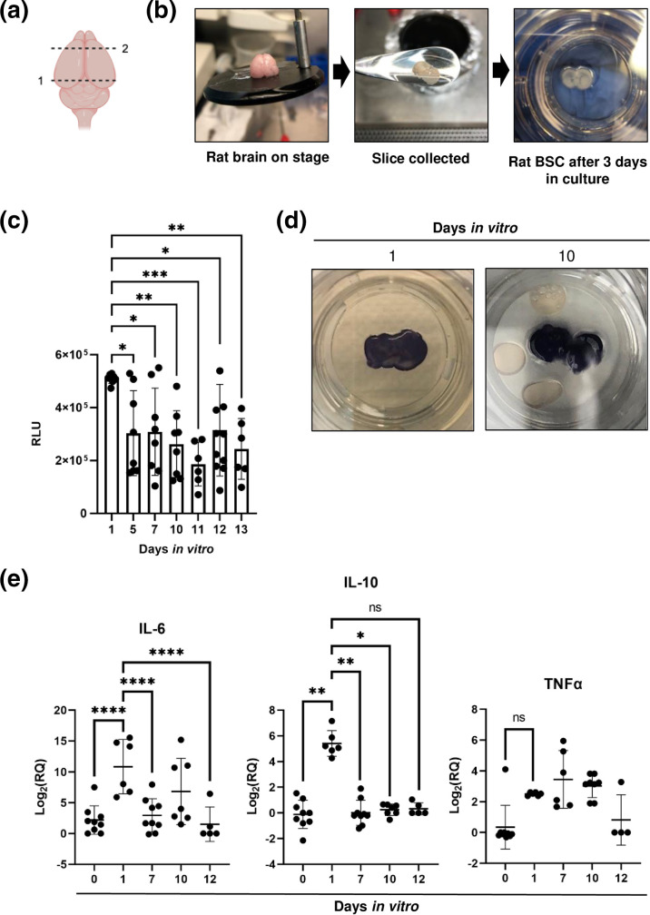
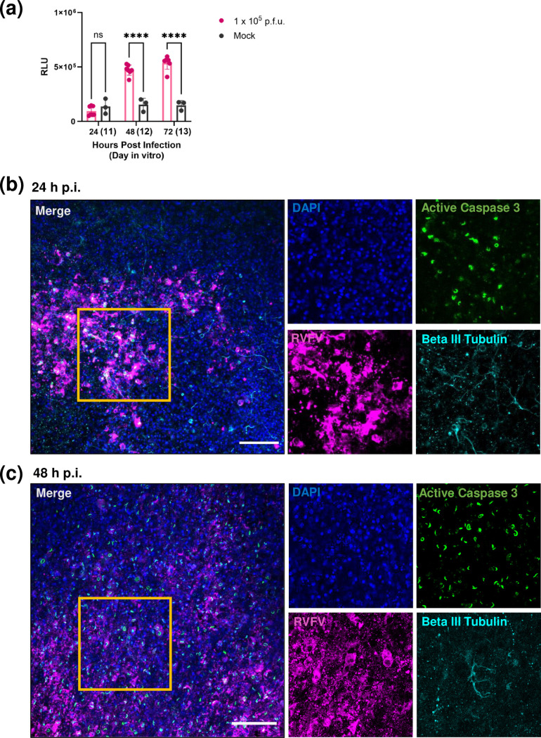
Rift Valley fever virus (RVFV) is an emerging arboviral disease with pandemic potential. While infection is often self-limiting, a subset of individuals may develop late-onset encephalitis, accounting for up to 20 % of severe cases. Importantly, individuals displaying neurologic disease have up to a 53 % case fatality rate, yet the neuropathogenesis of RVFV infection remains understudied. In this study, we evaluated whether ex vivo postnatal rat brain slice cultures (BSCs) could be used to evaluate RVFV infection in the central nervous system. BSCs mounted an inflammatory response after slicing, which resolved over time, and they were viable in culture for at least 12 days. Infection of rat BSCs with pathogenic RVFV strain ZH501 induced tissue damage and apoptosis over 48 h. Viral replication in BSCs reached up to 1×10⁷ p.f.u. equivalents/ml, depending on inoculation dose. Confocal immunofluorescent microscopy of cleared slices confirmed direct infection of neurons as well as activation of microglia and astrocytes. Further, RVFV-infected rat BSCs produced antiviral cytokines and chemokines, including MCP-1 and GRO/KC. This study demonstrates that rat BSCs support replication of RVFV for ex vivo studies of neuropathogenesis. This allows for continued and complementary investigation into RVFV infection in an ex vivo postnatal brain slice culture format.

Keywords: Rift Valley fever virus; brain slice culture; bunyavirus; ex vivo model; neurotropic virus; viral encephalitis.


Conflict of interest statement

The author(s) declare that there are no conflicts of interest.

Figures

Fig. 1.
Fig. 1.. Brain slices are isolated from postnatal rats and cultured ex vivo. (a) Schematic showing two cuts to whole brain (created in BioRender). Removal of the cerebellum and brain stem (cut 1) prior to mounting onto a vibratome stage. Removal of bi-lobular frontal cortex (cut 2). (b) Sequential, coronal brain sections were isolated from 6-day-old rat pups using a vibratome. Then, 400 µm sections were collected and seeded on PFTE membrane inserts. (c) Cellular cytotoxicity over time in relative luminescence units (RLU), quantified using LDH cytotoxicity assay (n>6 slices across three experiments). (d) Metabolic activity observed using MTT staining (purple=active, white=dead) in two representative slices from day 1 and day 10. (e) IL-10, IL-6 and TNFα expression in brain slices (n>5 slices across three experiments) quantified by delta-delta Ct fold change (beta-actin) normalized to day 0 BSC. Error bars represent sem. Statistical analysis performed using two-way ANOVA. *P < 0.05; **P < 0.01; ***P < 0.001; ****P<0.0001; ns=not significant.
Fig. 2.
Fig. 2.. RVFV infects, replicates, and spreads in ex vivo rat BSCs. Rat BSCs (n=5 per timepoint) were inoculated with 1×10⁵, 1×10⁴ or 1×10³ p.f.u. of pathogenic RVFV (strain ZH501) or mock infected and viral RNA (a) or infectious virus (b) was quantified at 24, 48 and 72 h p.i. (c) Mock or RVFV-infected slices at 24 h p.i. or 48 h p.i. were stained with anti-RVFV-N (magenta) and counterstained with DAPI (blue). Slices were cleared and imaged at 2.5× magnification. Scale bar=100 µm. Images are representative of five slices imaged per time point.
Fig. 3.
Fig. 3.. Rift Valley fever virus infects neurons and induces microglial changes in rat BSC. (a–c) Slices were fixed overnight with 4 % paraformaldehyde at different timepoints following infection with 1×10⁵ p.f.u. pathogenic RVFV (strain ZH501) or mock-infected control. Whole slices were stained with anti-RVFV-N (magenta), anti-IBA-1 (white), and anti-Beta III Tubulin (cyan), and counterstained with DAPI (blue). Slices were cleared, mounted to slides, and imaged at 20× on a Nikon A1 confocal across 40 µm z-plane. Yellow boxes over merged image are areas which are shown as single colour channels on the right. White arrow=co-localization of beta-III tubulin and RVFV-N. Yellow arrow=IBA-1+ microglia. Images represent maximum intensity projection. Scale bar=100 µm.
Fig. 4.
Fig. 4.. Rift Valley fever virus infection of rat BSCs promotes astrocyte activation. (a–c) Slices were fixed overnight with 4 % paraformaldehyde at different timepoints following infection with 1×10⁵ p.f.u. pathogenic RVFV (strain ZH501) or uninfected control. Whole slices were stained with anti-RVFV-N (magenta), anti-NeuN (green), and anti-GFAP (cyan), and counterstained with DAPI (blue). Slices were cleared, mounted to slides, and imaged at 20× on a Nikon A1 confocal across 40 µm z-plane. Yellow boxes over merged images are areas which are shown as single-colour channels on the right. White arrow=co-localization of NeuN and RVFV-N. Yellow arrow=GFAP+astrocyte. Images represent maximum intensity projection. Scale bar=100 µm.
Fig. 5.
Fig. 5.. Rift Valley fever virus infection induces tissue damage and the activation of apoptosis in ex vivo rat BSC. (a) Cellular cytotoxicity over time in relative luminescence units (RLU) between mock and slices infected with 1×10⁵ p.f.u. pathogenic RVFV (strain ZH501), quantified using LDH Glo Cytotoxicity Assay (n=5 infected slices and three mock slices per timepoint). Error bars represent sem. Statistical analysis performed using two-way ANOVA. *P < 0.05; **P < 0.01; ***P < 0.001; ****P<0.0001; ns=not significant. (b, c) Slices were fixed overnight with 4% paraformaldehyde at 24 and 48 h p.i. following infection with 1×10⁵ p.f.u. pathogenic RVFV (strain ZH501). Whole slices were stained with anti-RVFV-N (magenta), anti-Active Caspase 3 (green), and anti-Beta III tubulin (cyan), and counterstained with DAPI (blue). Slices were cleared and mounted to slides, then imaged at 20× on a Nikon A1 confocal across 40 µm z-plane. Yellow boxes over merged image are areas which are shown as single colour channels on the right. Images represent maximum intensity projection. Scale bar=100 µm.
Fig. 6.
Fig. 6.. Antiviral cytokines and chemokines are expressed during RVFV infection of ex vivo rat brain slices. (a, b)Rat BSC were infected with 1×10⁵ p.f.u. pathogenic RVFV (strain ZH501) or mock infected and RNA was isolated at 24, 48 or 72 h p.i. from the whole slice. Mock data represents mock-infected slices from each timepoint, and gene-expression data was pooled. Gene expression was quantified by RT-qPCR using delta-delta Ct and normalized to the variation of the amount of beta-actin within each slice. Data collected from three independent experiments. Fold changes are relative to a 0 h mock-infected control slice. Error bars represent sem. Statistical analysis performed using two-way ANOVA. *P < 0.05; **P < 0.01; ***P < 0.001; ****P<0.0001; ns=not significant.

Similar articles

Cited by

References

    1. Hartman A. Rift Valley fever. Clin Lab Med. 2017;37:285–301. doi: 10.1016/j.cll.2017.01.004. - DOI - PMC - PubMed
    1. Al-Hazmi M, Ayoola EA, Abdurahman M, Banzal S, Ashraf J, et al. Epidemic Rift Valley fever in Saudi Arabia: a clinical study of severe illness in humans. Clin Infect Dis. 2003;36:245–252. doi: 10.1086/345671. - DOI - PubMed
    1. Mohamed M, Mosha F, Mghamba J, Zaki SR, Shieh W-J, et al. Epidemiologic and clinical aspects of a Rift Valley fever outbreak in humans in Tanzania, 2007. Am J Trop Med Hyg. 2010;83:22–27. doi: 10.4269/ajtmh.2010.09-0318. - DOI - PMC - PubMed
    1. Bird BH, Ksiazek TG, Nichol ST, Maclachlan NJ. Rift Valley fever virus. J Am Vet Med Assoc. 2009;234:883–893. doi: 10.2460/javma.234.7.883. - DOI - PubMed
    1. Madani TA, Al-Mazrou YY, Al-Jeffri MH, Mishkhas AA, Al-Rabeah AM, et al. Rift Valley fever epidemic in Saudi Arabia: epidemiological, clinical, and laboratory characteristics. Clin Infect Dis. 2003;37:1084–1092. doi: 10.1086/378747. - DOI - PubMed

Publication types

LinkOut - more resources