. 2024 Apr 2;121(14):e2309000121.
doi: 10.1073/pnas.2309000121. Epub 2024 Mar 28.

Brainstem depolarization-induced lethal apnea associated with gain-of-function SCN1AL263V is prevented by sodium channel blockade

Affiliations

Brainstem depolarization-induced lethal apnea associated with gain-of-function SCN1AL263V is prevented by sodium channel blockade

Nico A Jansen et al. Proc Natl Acad Sci U S A. .

Abstract

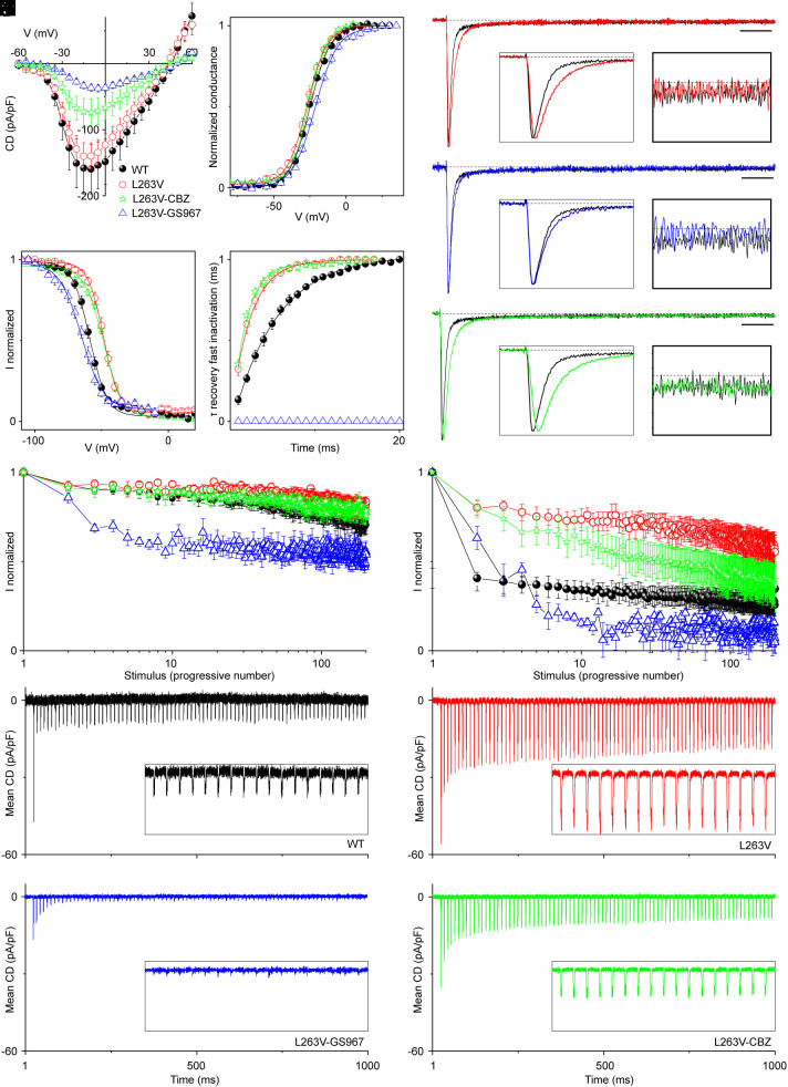
Apneic events are frightening but largely benign events that often occur in infants. Here, we report apparent life-threatening apneic events in an infant with the homozygous SCN1AL263V missense mutation, which causes familial hemiplegic migraine type 3 in heterozygous family members, in the absence of epilepsy. Observations consistent with the events in the infant were made in an Scn1aL263V knock-in mouse model, in which apnea was preceded by a large brainstem DC-shift, indicative of profound brainstem depolarization. The L263V mutation caused gain of NaV1.1 function effects in transfected HEK293 cells. Sodium channel blockade mitigated the gain-of-function characteristics, rescued lethal apnea in Scn1aL263V mice, and decreased the frequency of severe apneic events in the patient. Hence, this study shows that SCN1AL263V can cause life-threatening apneic events, which in a mouse model were caused by profound brainstem depolarization. In addition to being potentially relevant to sudden infant death syndrome pathophysiology, these data indicate that sodium channel blockers may be considered therapeutic for apneic events in patients with these and other gain-of-function SCN1A mutations.

Keywords: apnea; brainstem; sodium channel; sudden death.


Conflict of interest statement

Competing interests statement:The authors declare no competing interest.

Figures

Fig. 1.
Fig. 1.
A patient with homozygosity for the SCN1AL263V mutation in an FHM3 family. (A) Electropherograms showing single or double copy of the C→G nucleotide change in codon 263 of exon 6 of the SCN1A gene, respectively in heterozygous and homozygous carriers. (B) Pedigree of the patient’s family, showing heterozygous (half-filled) and homozygous (proband (indicated by the arrow); filled) carriers of the SCN1AL263V mutation, as well as wild-type (empty) and nontested (question mark) family members. (C) Axial brain 3T MR images obtained from the proband at 17 mo following an episode of severe apnea and cardiac arrest requiring cardiopulmonary resuscitation, showing T2-weighted hyperintensity of the basal ganglia bilaterally (Top) with areas of restricted diffusion (Bottom, arrows), consistent with hypoxic-ischemic brain injury. (D) Frequency (black and gray) and duration (red) of apneic events in the proband, with particularly severe events indicated by arrows at the top (ALTE = apparent life-threatening event; CA = cardiac arrest). Note that event duration was not affected by levetiracetam (LEV; 40 mg/kg/day), but decreased following addition of carbamazepine (CBZ; 10 mg/kg/day). Although an increase in minor events (lasting 5 to 10 s) was observed (gray line), this may reflect an increased awareness of the caregiver and/or physician for such events, which was nevertheless followed by an increase in CBZ dosage to 17 mg/kg/day at 33 mo.
Fig. 2.
Fig. 2.
Example of brain and cardiorespiratory recordings during spontaneous sudden death in a heterozygous Scn1aL263V mouse. Note that the sudden onset of apnea coincided with transient bradycardia, and preceded suppression of electrocorticogram activity (cortex AC).
Fig. 3.
Fig. 3.
Lethal sudden apnea in heterozygous Scn1aL263V mice is preceded by a DC-shift in the medulla. (A) Spontaneous sudden apnea and bradycardia follow soon after onset of a DC-shift in the ventrolateral medulla (dashed line, corresponding to arrowheads in Insets) in an Scn1aL263V mouse. (B) Similar dynamics were observed in recordings from all Scn1aL263V mice (n = 13 with respiratory recordings, n = 6 with ECG), here aligned at onset of the medullary DC-shift (dashed line). When compared to baseline (I), respiratory rate was significantly increased prior to the DC-shift (II), while both respiratory and heart rate were decreased immediately after the DC-shift (III; *P < 0.05, **P < 0.005, repeated measures ANOVA with Dunnett’s test). (C) Timing of arrest of brain and cardiorespiratory activity with respect to the medullary DC-shift. A consistent sequence of apnea, EEG suppression, cortical AD and cardiac arrest was observed in all animals.
Fig. 4.
Fig. 4.
Sudden apnea in heterozygous Scn1aL263V mice is induced by a profound brainstem depolarization. (A) Example of spontaneous and near-simultaneous depolarizations in the pons and medulla of an Scn1aL263V mouse. (B) Brainstem DC-shifts (onset indicated by dashed line, arrowhead in the Inset) following stimulation of the inferior colliculus (Stim) preceded medullary and cortical local PO2 drops. Note that, on the other hand, the cortical DC-shift followed cortical hypoxia, indicative of an AD (asterisk). (C) Following stimulation of the inferior colliculus, medullary hypoxia (PO2 < 10 mmHg) consistently preceded cortical hypoxia. (D) Local PO2 consistently decreased faster in the medulla when compared to the cortex. (E) Lethality following stimulation of the inferior colliculus could be prevented by timely respiratory resuscitation (*P = 0.010; Mann–Whitney test). Resuscitation was also successful during witnessed spontaneous events in two Scn1aL263V mice (red).
Fig. 5.
Fig. 5.
Profound brainstem depolarization in heterozygous Scn1aL263V mice preceding spontaneous fatal apnea. (A) Average DC-signal in the brainstem ventrolateral medulla synchronized at onset of the DC-shift (dashed line at time = 0 s). Timing of DC-shift onset in the pons (n = 5, blue) and contralateral medulla (n = 6, red) is indicated at the Top. Note that an increase in medullary MUA accompanies the negative DC-shift, and is suppressed following the event. (B) Average respiratory rate and medullary MUA in Scn1aL263V mice (n = 5) at the time of the medullary DC-shift (dashed line). Note that average MUA is slightly different since only animals with high-quality respiratory recordings are included.
Fig. 6.
Fig. 6.
Functional modifications induced by L263V on hNav1.1 and effects of sodium channel blockers GS967 and CBZ. (A) Current density-voltage plots for tsA-201 cells transfected with hNav1.1-WT (black), hNav1.1-L263V (red), hNav1.1-L263V recorded in the presence of 15 µM of CBZ (green) and hNav1.1-L263V recorded in the presence of 5 µM GS967 (blue). (B) Mean voltage dependence of activation, lines are Boltzmann fits. GS967 induced a 6.1-mV positive shift of the L263V curve (P = 0.002), mainly caused by the decrease of the steepness of the curve (20% increase of L263V slope factor, ka, P = 0.007). No statistically significant difference was present between the activation curves of WT, L263V, and L263V-CBZ. (C) Mean voltage dependence of fast inactivation, lines are Boltzmann fits. (D) Recovery of fast inactivation at −80 mV. (E and G) Comparison of average normalized currents elicited by depolarizing steps to −10 mV for hNav1.1-WT and hNav1.1-L263V (E), hNav1.1 and hNav1.1-L263V-GS967 (F); hNav1.1 and hNav1.1-L263V-CBZ (G); calibration bar 5 ms. The Left Insets show the first 6 ms of the current traces to compare the current decay, the Right Insets show the traces between 73.5 to 78.5 ms to compare INaP. (H and I) Use dependence (current normalized to the first stimulus in the train) induced by trains of 2-ms depolarizing steps to 0 mV from the holding potential of −70 mV at a frequency of 10 Hz (H) and 100 Hz (I) for hNav1.1-WT, hNav1.1-L263V, hNav1.1-L263V-GS967 and hNav1.1-L263V-CBZ. (JM) Action Na+ currents expressed as mean current density recorded using as voltage stimulus the action potential discharge of a GABAergic fast spiking neuron for hNav1.1-WT (J), hNav1.1-L263V (K), hNav1.1-L263V-GS967 (L) and hNav1.1-L263V-CBZ (M). Data points are displayed as mean ± SEM. See SI Appendix, Figs. S3 and S4 and Table S1 for statistical comparisons.
Fig. 7.
Fig. 7.
Sodium channel blockers prevent lethal apnea in heterozygous Scn1aL263V mice. (A) Survival of Scn1aL263V mice is improved by chow compounded by GS967 (8 mg/kg chow; red; P < 0.0001, Mantel-Cox test). In a separate group, withdrawal from GS967 at P42 (blue, arrow) reinstated the sudden death phenotype. (B) Stimulation of the inferior colliculus was less likely fatal in mice maintained on GS967-compounded, when compared to normal, chow (P < 0.0001, Fisher’s exact test). (C) Intraperitoneal pretreatment with GS967 or CBZ prevented death following stimulation, when compared to vehicle (VEH; P < 0.0001, Fisher’s exact test), whereas MK-801 did not increase survival at the two doses tested (P > 0.99, Fisher’s exact test).

References

    1. DiMario F. J. Jr., Breath-holding spells in childhood. Am. J. Dis. Child. 146, 125–131 (1992). - PubMed
    1. Kahn A., et al. , Sleep and cardiorespiratory characteristics of infant victims of sudden death: A prospective case-control study. Sleep 15, 287–292 (1992). - PubMed
    1. Thach B. T., Potential central nervous system involvement in sudden unexpected infant deaths and the sudden infant death syndrome. Compr. Physiol. 5, 1061–1068 (2015). - PubMed
    1. Koh H. Y., et al. , Genetic determinants of sudden unexpected death in pediatrics. Genet. Med. 24, 839–850 (2022). - PMC - PubMed
    1. Brownstein C. A., et al. , SCN1A variants associated with sudden infant death syndrome. Epilepsia 59, e56–e62 (2018). - PMC - PubMed

Substances

LinkOut - more resources