. 2024 Aug;13(21):e2304338.
doi: 10.1002/adhm.202304338. Epub 2024 Apr 8.

Sided Stimulation of Endothelial Cells Modulates Neutrophil Trafficking in an In Vitro Sepsis Model

Affiliations

Sided Stimulation of Endothelial Cells Modulates Neutrophil Trafficking in an In Vitro Sepsis Model

Danial Ahmad et al. Adv Healthc Mater. 2024 Aug.

Abstract

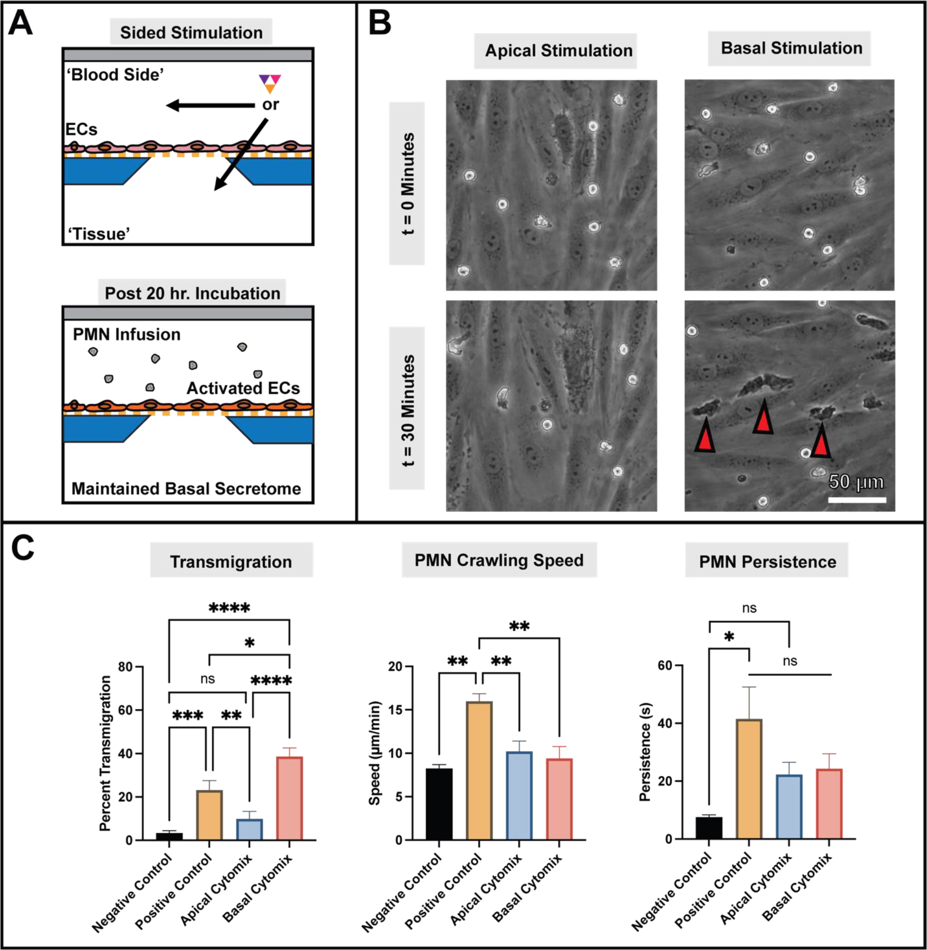
While the role of dysregulated polymorphonuclear leukocyte (PMN) transmigration in septic mediated tissue damage is well documented, strategies to mitigate aberrant transmigration across endothelium have yet to yield viable therapeutics. Recently, microphysiological systems (MPS) have emerged as novel in vitro mimetics that facilitate the development of human models of disease. With this advancement, aspects of endothelial physiology that are difficult to assess with other models can be directly probed. In this study, the role of endothelial cell (EC) apicobasal polarity on leukocyte trafficking response is evaluated with the µSiM-MVM (microphysiological system enabled by a silicon membrane - microvascular mimetic). Here, ECs are stimulated either apically or basally with a cytokine cocktail to model a septic-like challenge before introducing healthy donor PMNs into the device. Basally oriented stimulation generated a stronger PMN transmigratory response versus apical stimulation. Importantly, healthy PMNs are unable to migrate towards a bacterial peptide chemoattractant when ECs are apically stimulated, which mimics the attenuated PMN chemotaxis seen in sepsis. Escalating the apical inflammatory stimulus by a factor of five is necessary to elicit high PMN transmigration levels across endothelium. These results demonstrate that EC apicobasal polarity modulates PMN transmigratory behavior and provides insight into the mechanisms underlying sepsis.

Keywords: apicobasal polarity; membranes; microphysiological systems; neutrophil trafficking; sepsis; vascular barriers.


Conflict of interest statement

Conflict of Interest

J.L.M. cofounded SiMPore and holds an equity interest in the company. SiMPore is a company that focuses on commercializing ultrathin silicon-based technologies for a variety of purposes including the devices used in this publication.

Figures

Figure 1.
Figure 1.. HUVECs are responsive to a septic-like cytokine cocktail when stimulated either apically or basally.
(A) The μSiM-MVM device features two flow channels that are separated by a porous membrane interface, facilitating stimulatory access to both the apical and basal surfaces on HUVECs mimicking ‘blood sided’ or ‘tissue sided’ inflammation respectively. For these studies, an equimolar combination of TNF-α, IL-1β, and IFN-γ (‘cytomix’) was used to mimic septic-like and tissue-oriented inflammation. (B) The incorporation of 50 pg/mL cytomix, regardless of stimulation orientation, activated HUVECs as measured by increased expression of ICAM-1 (AF488). Notably, the bacterial peptide and PMN chemoattractant fMLP does not activate HUVECs upon a 30 minute exposure with 10 nM concentration. (C) When evaluating z-axis organization of ICAM-1, basally stimulated HUVECs generated robust luminal ICAM-1 projections or “hill-like” formations while apically stimulated HUVECs appeared morphologically flatter. Experiments were performed in triplicate with sequential passages of HUVECs.
Figure 2.
Figure 2.. Activated endothelium alone, regardless of stimulation orientation, is insufficient for promoting PMN transmigration across endothelium.
(A) To mechanistically isolate activated HUVEC monolayer contributions to PMN transmigration, μSiM-MVM devices with HUVECs were incubated with cytomix in either channel. The devices were then flushed with fresh media after 20 hours of incubation to remove any chemokine gradients. Afterwards, PMNs were incorporated and monitored in phase contrast. (B) PMNs were separately incorporated into μSiM-MVM devices at a concentration of 3 million cells/mL. Conditions assessed include no stimulation (negative control), a basally oriented 10 nM fMLP gradient (positive control), and directionally stimulated endothelium with flushed channels (apical and basal stimulation). Phase contrast time-lapse recordings were collected immediately upon PMN introduction for thirty minutes of coculture incubation, with experiment start (t = 0 minutes, first row) and end (t = 30 minutes, second row) presented as images. Red arrows highlight occurrences of PMN transmigration, and all images are representative of triplicate studies. For all assessed studies, only PMNs stimulated with fMLP gradients actively transmigrated across the endothelium. (C) Average data tabulated from time-lapse studies demonstrates the lack of PMN transmigration in either sided stimulation group, with both presenting statistically similar results to the negative control study (< 3.5% population transmigration). In contrast, the positive control study displays significantly higher transmigration results. Despite the lack of transmigration, PMNs crawl faster on stimulated endothelium with statistically similar speeds to fMLP stimulated PMNs. PMN persistence appears to depend on the sidedness of the prior EC stimulation, with higher values on basally stimulated ECs and lower on apical. PMN persistence was found to be statistically similar when comparing the fMLP and basal stimulation groups, as well as the negative control and apical stimulation groups. Statistics: Sample size n = 3, * = P < 0.05, ** = P < 0.001, *** = P < 0.0001. Comparisons made with a one way ANOVA and error bars represent standard error of mean.
Figure 3.
Figure 3.. Maintaining EC secretome environment post 20 hour sided cytomix incubation elicits differential PMN transmigratory behavior.
(A) Experimental protocol followed the same steps as the previous experiments, but without flushing the basal channel prior to the introduction of PMNs. (B) PMNs incorporated into μSIM-MVM devices (3 million cells/mL) were observed in phase contrast time-lapse recordings over thirty minutes. Representative images shown. The red arrows highlight occurrences of PMN transmigration. When the secretome gradient is maintained, PMNs begin to exhibit transmigratory behavior on the basally stimulated endothelium while little transmigration is observed in apically stimulated devices. (C) In this instance, apically stimulated devices elicited statistically similar PMN transmigration rates as negative controls, while basally stimulated devices exhibited significantly more PMN transmigration than positive controls. Despite this, PMN speeds and persistence were lower in the sided stimulation groups, with results matching negative controls. PMNs crawled fastest when stimulated with fMLP. Statistics: Sample size n = 3, * = P < 0.05, ** = P < 0.001, *** = P < 0.0001, **** = P < 0.00001. Comparisons made with a one way ANOVA and error bars represent standard error of mean.
Figure 4.
Figure 4.. Cytomix components were individually assessed to determine if any single cytokine contributed greatly to the high levels of PMN transmigration seen with basally stimulated ECs.
(A) A series of μSiM-MVM devices with HUVEC monolayers were basally stimulated with 50 pg/mL of each cytomix component individually. After 20 hours of incubation, PMNs were seeded on top of ECs at a concentration of 3 million cells/mL, then observed in phase contrast time lapse recordings for 30 minutes. (B) PMN transmigration and persistence were similar across all tested groups, indicating no single cytokine contributes disproportionately to PMN trafficking. PMN speed was different, however, as PMNs crawled fastest on IL-1β stimulated ECs. (C - D) The analytical methods used in this study can assess PMN motility parameters (speed and persistence) both before and after transmigration. With single cytokine studies, transmigration typically has no effect on crawling speed or persistence except in the case of IFN-γ, where PMNs became less persistent after transmigration. Statistics: Sample size n = 3, * = P < 0.05, *** = P < 0.0001, ns = not significant. Comparisons made with a one way ANOVA and error bars represent standard error of mean.
Figure 5.
Figure 5.. Increasing stimulus levels results in higher levels of transmigration for both apical and basal stimulus.
(A) Experiments were performed with increased (250 pg/mL) levels of cytomix to assess if changes would occur in PMN trafficking behavior. Regardless of stimulation orientation, 250 pg/mL cytomix elicits significantly greater PMN transmigration than observed at 50 pg/mL. At 250 pg/mL, ECs that are basally stimulated with cytomix facilitate higher PMN transmigration than all other tested conditions, with average transmigration exceeding 80%. Despite this, PMN motility parameters did not change significantly at a high cytomix concentration when compared to the low concentration studies. (B) In all experimental scenarios, transmigrated PMNs also crawled at a similar rate to non-transmigrated cells. (C) PMNs in the higher concentration cytomix studies were significantly more persistent when migrating on the luminal surface of the monolayer, but persistence for abluminal crawling was unchanged. Statistics: Sample size n = 3 per group, * = P < 0.05, ns = not significant. Comparisons made with a one way ANOVA and error bars represent standard error of mean.
Figure 6.
Figure 6.. PMNs exhibit attenuated chemotaxis towards fMLP gradients when either ECs or PMNs
Experiments were performed to assess the impact of cytomix on PMN homing capabilities towards basally oriented 10 nM fMLP gradients. A normal positive control study was compared against similar studies where either ECs were apically stimulated for 20 hours with cytomix, or PMNs stimulated for 15 minutes prior to introduction into the device. In all cases, a 10 nM fMLP gradient was used to mimic a bacterial stimulus originating from the tissue. (B) The addition of either cytomix stimulated ECs or pre-stimulated PMNs resulted in decreased PMN transmigration across the HUVEC endothelium, even when fMLP gradients were present. The positive control study (fMLP only, normal ECs and PMNs) displayed significantly higher transmigration than all other conditions, with the cytomix + fMLP studies displaying similar transmigration levels to the negative control and apical cytomix group. In all cases, PMNs were slower than PMNs in the positive control group, with the exception of cytomix stimulated PMNs. The presence of fMLP, regardless of cytomix stimulation, increased PMN persistence for all studied groups. Statistics: Sample size n = 3, * = P < 0.05, ** = P < 0.001, *** = P < 0.0001. Comparisons made with a one way ANOVA and error bars represent standard error of mean.

Similar articles

Cited by

References

    1. Nduka OO and Parrillo JE, Critical Care Clinics, 2009, 25, 677–702. - PubMed
    1. Rudd KE, Kissoon N, Limmathurotsakul D, Bory S, Mutahunga B, Seymour CW, Angus DC and West TE, Critical Care, 2018, 22. - PMC - PubMed
    1. Martin GS, Mannino DM, Eaton S and Moss M, N Engl J Med, 2003, 348, 1546–1554. - PubMed
    1. Rubens M, Saxena A, Ramamoorthy V, Das S, Khera R, Hong J, Armaignac D, Veledar E, Nasir K and Gidel L, J Intensive Care Med, 2020, 35, 858–868. - PubMed
    1. Fink MP and Warren HS, Nature Reviews Drug Discovery, 2014, 13, 741–758. - PubMed

MeSH terms