. 2024 Mar 28;15(1):2742.
doi: 10.1038/s41467-024-45594-4.

Comprehensive mutational scanning of EGFR reveals TKI sensitivities of extracellular domain mutants

Affiliations

Comprehensive mutational scanning of EGFR reveals TKI sensitivities of extracellular domain mutants

Tikvah K Hayes et al. Nat Commun. .

Erratum in

Abstract

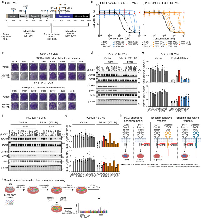
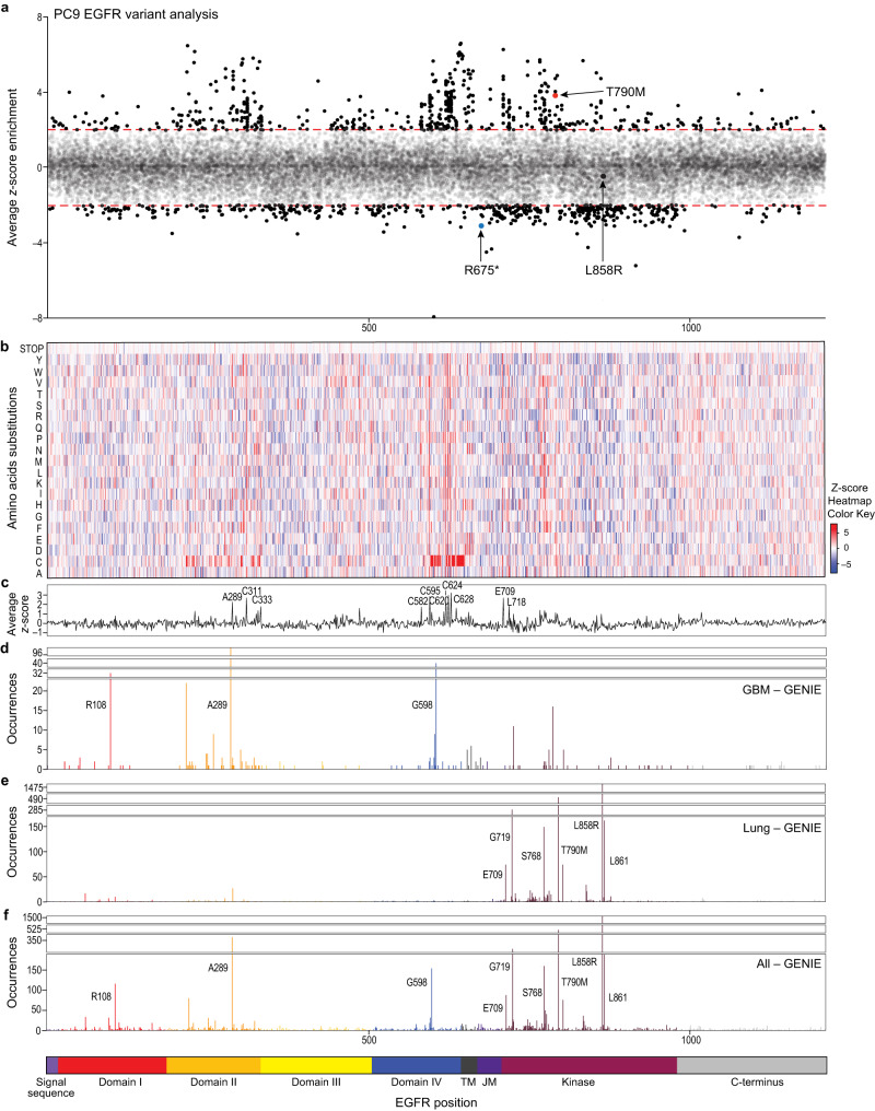
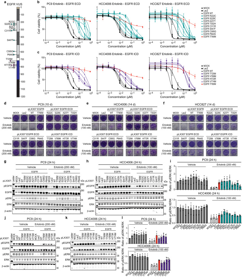
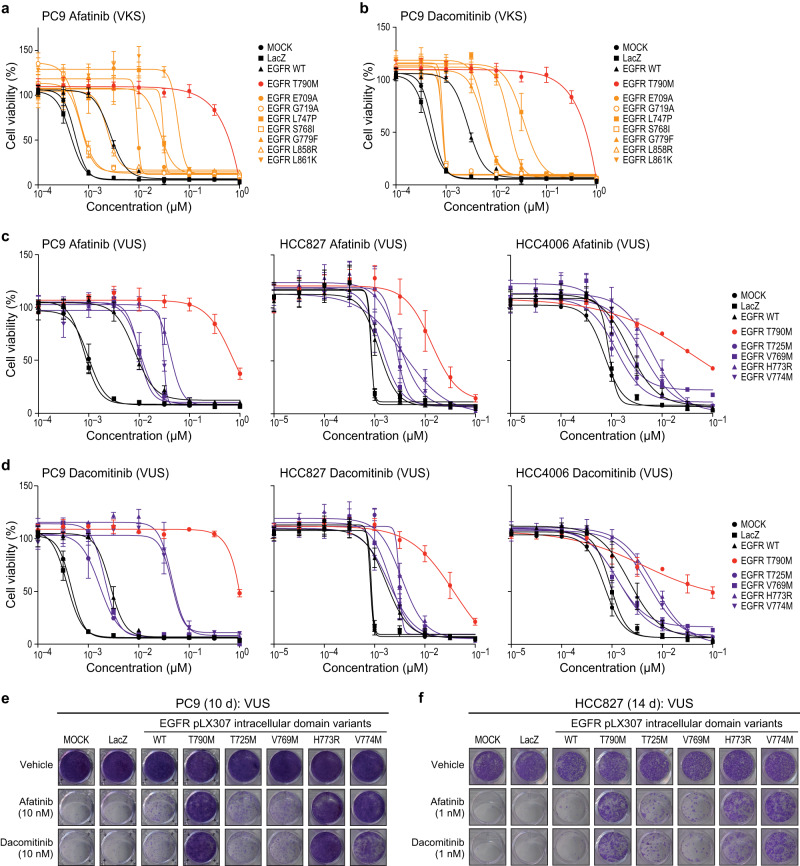
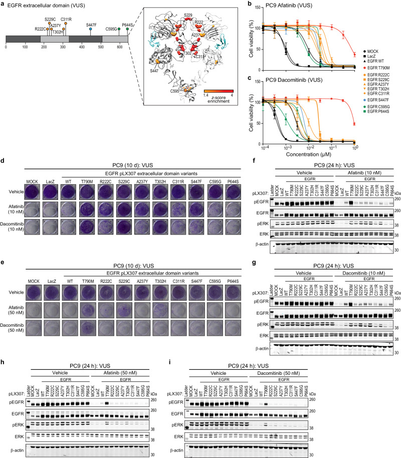
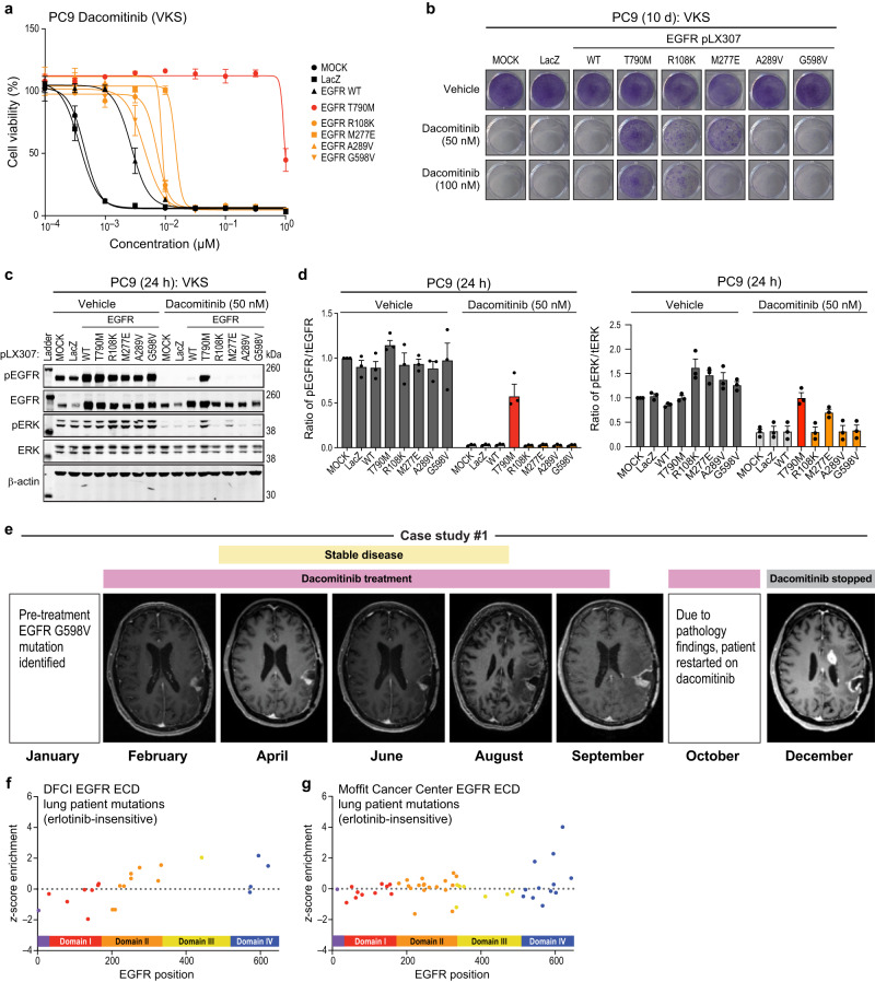
The epidermal growth factor receptor, EGFR, is frequently activated in lung cancer and glioblastoma by genomic alterations including missense mutations. The different mutation spectra in these diseases are reflected in divergent responses to EGFR inhibition: significant patient benefit in lung cancer, but limited in glioblastoma. Here, we report a comprehensive mutational analysis of EGFR function. We perform saturation mutagenesis of EGFR and assess function of ~22,500 variants in a human EGFR-dependent lung cancer cell line. This approach reveals enrichment of erlotinib-insensitive variants of known and unknown significance in the dimerization, transmembrane, and kinase domains. Multiple EGFR extracellular domain variants, not associated with approved targeted therapies, are sensitive to afatinib and dacomitinib in vitro. Two glioblastoma patients with somatic EGFR G598V dimerization domain mutations show responses to dacomitinib treatment followed by within-pathway resistance mutation in one case. In summary, this comprehensive screen expands the landscape of functional EGFR variants and suggests broader clinical investigation of EGFR inhibition for cancers harboring extracellular domain mutations.


Conflict of interest statement

M.M. receives research support from Bayer Pharmaceuticals and Janssen. M.M. is a consultant for and equity holder in DelveBio, Interline and Isabl, and holds patents licensed to Bayer and LabCorp. C.M.J. is a full-time employee of and equity holder in Novartis. D.P.C. consults for the Massachusetts Institute of Technology, Advise Connect Inspire, German Accelerator, Lilly, GlaxoSmithKline, Incephalo, Boston Pharmaceuticals, Servier, Boston Scientific and Pyramid Biosciences (equity interest) for advisory input. He has also received financial compensation and travel reimbursement from Merck for invited lectures, and from the US NIH and DOD for clinical trial and grant review. F.P. is a full-time employee of Merck Research Laboratories. A.D.C. receives research support from Bayer Pharmaceuticals. D.E.R. receives research funding from members of the Functional Genomics Consortium (Abbvie, Bristol-Myers Squibb, Jannsen, Merck, Vir), and is a director of Addgene, Inc. A.S.C. is a full-time employee of and equity holder in Alterome Therapeutics. J.D. is a consultant for Novartis, Amgen, Janssen and Ono Therapeutics. N.S.P. is a full-time employee of Aera Therapeutics. The remaining authors declare no competing interests. J.K.H. serves as a consultant for Jackson Laboratory for Genomic Medicine.

Figures

Fig. 1
Fig. 1. Validation of VKS in PC9 oncogene addiction model and screening schematic.
a EGFR domain schematic showing tested variants of known significance (VKS). b PC9 cell lines expressing either LacZ, EGFR WT, EGFR T790M, EGFR extracellular domain variants (R108K, M277E, A289V, or G598V), or EGFR intracellular domain variants (E709A, G719A, L747P, S768I, G779F, L858R or L861K), after 144 h treatment with increasing doses of erlotinib (normalized to vehicle control). A representative experiment is shown, remaining biological replicates are located in Source Data (N = 3). Data are presented as mean values ± SD. c Colony formation assay in PC9 cell lines expressing either LacZ, EGFR WT, EGFR T790M, EGFR extracellular domain variants (R108K, M277E, A289V, or G598V), or EGFR intracellular domain variants (E709A, G719A, L747P, S768I, G779F, L858R or L861K) after a 10 d of treatment with either vehicle (DMSO) or 200 nM erlotinib. A representative experiment is shown (N = 3). dg Representative immunoblots displaying the effect of 200 nM erlotinib after 24 h treatment on PC9 cell lines expressing either LacZ, EGFR WT, EGFR T790M, EGFR extracellular domain variants (R108K, M277E, A289V, or G598V) (d, e), or EGFR intracellular domain variants (E709A, G719A, L747P, S768I, G779F, L858R or L861K) (f, g) on the levels of both phosphorylated EGFR and ERK and total EGFR and ERK. β-actin immunoblotting was used to determine equivalent loading. Data are presented as mean values ± SEM of biological replicates (N = 3) (e, g). h PC9 oncogene addiction model. i Screening schematic and timeline.
Fig. 2
Fig. 2. Systematic identification of erlotinib-insensitive EGFR variants using deep mutational scanning.
af PC9 cell line stably expressing an EGFR missense variant library was treated for 10 d with 200 nM erlotinib. Alignment of EGFR variant library analysis by z-score enrichment of position (a) or amino acid substitution (b), average z-score by position (c), and EGFR patient observed mutations in glioblastoma (d), lung (e), and all cancers (f) (GENIE). For the EGFR schematic below, TM (transmembrane) and JM (juxtamembrane).
Fig. 3
Fig. 3. Characterization of EGFR variants of unknown significance (VUS) in lung cancer models of EGFR addiction.
a EGFR domain schematic showing tested variants of unknown significance (VUS). b, c PC9, HCC4006, or HCC827 cell lines expressing either LacZ, EGFR WT, EGFR T790M, EGFR extracellular domain variants (R222C, S229C, A237Y, T302H, C311R, S447F, C595G, or P644S) (b), or EGFR intracellular domain variants (T725M, V769M, H773R, or V774M) (c) after 144 h treatment with increasing doses of erlotinib (normalized to vehicle control). A representative experiment is shown, remaining biological replicates are located in Source Data (N = 3). Data are presented as mean values +/- SD (b, c). d-f Colony formation with PC9 EGFR mutants and controls as in (b) and (c) after 10 d (PC9) or 14 d (HCC4006 and HCC827) of treatment with either vehicle (DMSO), 100 nM erlotinib (HCC4006 or HCC827), or 200 nM erlotinib (PC9). A representative experiment is shown (N = 3). g–l Representative immunoblots displaying the effect of either 200 nM (PC9) or 100 nM (HCC4006) erlotinib after 24 h treatment of cell lines expressing either LacZ, EGFR WT, EGFR T790M, EGFR extracellular domain variants (R222C, S229C, A237Y, T302H, C311R, S447F, C595G, or P644S) (g, h, i), or EGFR intracellular domain variants (T725M, V769M, H773R, and V774M) (j, k, l) on the levels of both phosphorylated EGFR and ERK and total EGFR and ERK. β-actin immunoblotting was used to determine equivalent loading. Data are presented as mean values ± SEM of biological replicates (PC9, N = 4; HCC4006, N = 3) (i, l).
Fig. 4
Fig. 4. The effects of osimertinib treatment on EGFR intracellular domain variants.
a EGFR domain schematic and ribbon structure of both EGFR intracellular domain VKS and VUS. b PC9 cell lines expressing either LacZ, EGFR WT, EGFR T790M (controls), or EGFR VKS intracellular domain variants after 144 h treatment with increasing doses of osimertinib. A representative experiment is shown, remaining biological replicates are located in Source Data (N = 3). Data are presented as mean values ± SD. c Colony formation with PC9 EGFR mutants and controls after 10 d of 10 nM osimertinib treatment. A representative experiment is shown (N = 3). d Representative immunoblots displaying the effect of 10 nM osimertinib after 24 h treatment on PC9 EGFR mutants and controls on the levels of phosphorylated EGFR and ERK and total EGFR and ERK. β-actin is the loading control. e Colony formation with PC9 EGFR mutants and controls after 10 d of 50 or 100 nM osimertinib treatment. A representative experiment is shown (N = 3). f, g Representative immunoblots displaying the effect of 100 (f) nM osimertinib after 24 h treatment on PC9 EGFR mutants and controls on the levels of phosphorylated EGFR and ERK and total EGFR and ERK. β-actin is the loading control. Total levels of phosphorylated-EGFR or ERK were normalized to total levels of EGFR or ERK. Presented data are mean values ± SEM of biological replicates (N = 3) (g). h PC9 cell lines expressing controls or EGFR VUS intracellular domain variants after 144 h treatment with increasing doses of osimertinib. A representative experiment is shown, remaining biological replicates are located in Source Data (N = 3). Data are presented as mean values ± SD. i, j Colony formation with PC9 EGFR mutants and controls after 10 d of 10, 50 or 100 nM osimertinib treatment. A representative experiment is shown (N = 3). km Representative immunoblots displaying the effect of 10 (k) or 100 (l) nM osimertinib after 24 h treatment on PC9 EGFR mutants and controls on the levels of phosphorylated EGFR and ERK and total EGFR and ERK. β-actin is the loading control. Total levels of phosphorylated-EGFR or ERK were normalized to total levels of EGFR or ERK. Presented data are mean values ± SEM of biological replicates (N = 3) (m).
Fig. 5
Fig. 5. EGFR intracellular domain variant sensitivity to afatinib and dacomitinib.
a, b PC9 cell lines expressing either LacZ, EGFR WT, EGFR T790M, or EGFR VKS intracellular domain variants (E709A, G719A, L747P, S768I, G779F, L858R or L861K) after 144 h treatment with increasing doses of afatinib (a) or dacominitib (b) in (normalized to vehicle control). A representative experiment is shown, remaining biological replicates are located in Source Data (N = 3). Data are presented as mean values ± SD. c, d PC9 cell lines expressing either LacZ, EGFR WT, EGFR T790M, EGFR extracellular domain variants (T725M, V769M, H773R, and V774M) after 144 h treatment with increasing doses of afatinib (c) or dacomitinib (d) (normalized to vehicle control). A representative experiment is shown, remaining biological replicates are located in Source Data (N = 3). Data are presented as mean values ± SD. ef Colony formation with either PC9 (e) or HCC827 (f) EGFR mutants and controls as in (c) and (d) after 10 or 14 d of treatment with either vehicle (DMSO), 1 nM, 10 nM,  of either afatinib or dacomitinib. A representative experiment is shown (N = 3).
Fig. 6
Fig. 6. EGFR extracellular domain variant sensitivity to afatinib and dacomitinib.
a Domain schematic and ribbon structure of EGFR extracellular domain variants of unknown significance (VUS). b, c PC9 cell lines expressing either LacZ, EGFR WT, EGFR T790M, EGFR extracellular domain variants (R222C, S229C, A237Y, T302H, C311R, S447F, C595G, or P644S) after 144 h treatment with increasing doses of afatinib (b) or dacomitinib (c) (normalized to vehicle control). A representative experiment is shown, remaining biological replicates are located in Source Data (N = 3). Data are presented as mean values ± SD. d, e Colony formation with PC9 EGFR mutants and controls as in (b) and (c) after 10 d of treatment with either vehicle (DMSO), 10 nM (d) or 50 nM (e) of either afatinib or dacomitinib. A representative experiment is shown (N = 3). fi Representative immunoblots displaying the effect of either afatinib (f, h) or dacominitib (g, i) after 24 h treatment on PC9 EGFR mutants and controls as in (b) and (c) on the levels of both phosphorylated EGFR and ERK and total EGFR and ERK. β-actin immunoblotting was used to determine equivalent loading.
Fig. 7
Fig. 7. Treatment of glioblastoma harboring EGFR G598V mutation with dacomitinib.
a PC9 cell lines expressing either LacZ, EGFR WT, EGFR T790M, EGFR VKS extracellular domain variants (R108K, M277E, A289V, or G598V) after 144 h treatment with increasing doses of dacomitinib (normalized to vehicle control). A representative experiment is shown, remaining biological replicates are located in Source Data (N = 3). Data are presented as mean values ± SD. b Colony formation with PC9 EGFR mutants and controls as in (a) after 10 d of treatment with either vehicle (DMSO), 50 nM, or 100 nM dacomitinib. A representative experiment is shown (N = 3). c, d Representative immunoblots displaying the effect of 10 nM dacomitinib after 24 h treatment on PC9 EGFR mutants and controls as in (a) on the levels of both phosphorylated EGFR and ERK and total EGFR and ERK. β-actin immunoblotting was used to determine equivalent loading (c). Total levels of phosphorylated-EGFR or ERK were normalized to total levels of either EGFR or ERK. Data are presented as mean values ± SEM of biological replicates (N = 3) (d). e Images of an EGFR G598V positive glioblastoma patient after treatment with dacomitinib. f, g EGFR extracellular domain mutations observed in lung cancer patients from Dana-Farber Cancer Institute (f) or Moffit Cancer Center (g) based on z-score enrichment from the screen.

References

    1. Brennan CW, et al. The somatic genomic landscape of glioblastoma. Cell. 2013;155:462–477. doi: 10.1016/j.cell.2013.09.034. - DOI - PMC - PubMed
    1. Cancer Genome Atlas Research, N. Comprehensive molecular profiling of lung adenocarcinoma. Nature. 2014;511:543–550. doi: 10.1038/nature13385. - DOI - PMC - PubMed
    1. Consortium, A. P. G. AACR Project GENIE: powering precision medicine through an international consortium. Cancer Discov. 2017;7:818–831. doi: 10.1158/2159-8290.CD-17-0151. - DOI - PMC - PubMed
    1. Lee JC, et al. Epidermal growth factor receptor activation in glioblastoma through novel missense mutations in the extracellular domain. PLoS Med. 2006;3:e485. doi: 10.1371/journal.pmed.0030485. - DOI - PMC - PubMed
    1. Lynch TJ, et al. Activating mutations in the epidermal growth factor receptor underlying responsiveness of non-small-cell lung cancer to gefitinib. N. Engl. J. Med. 2004;350:2129–2139. doi: 10.1056/NEJMoa040938. - DOI - PubMed

MeSH terms