. 2024 Mar 28;14(1):7427.
doi: 10.1038/s41598-024-57973-4.

Taurine reduces microglia activation in the brain of aged senescence-accelerated mice by increasing the level of TREM2

Affiliations

Taurine reduces microglia activation in the brain of aged senescence-accelerated mice by increasing the level of TREM2

Sharif Ahmed et al. Sci Rep. .

Abstract

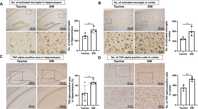
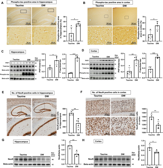
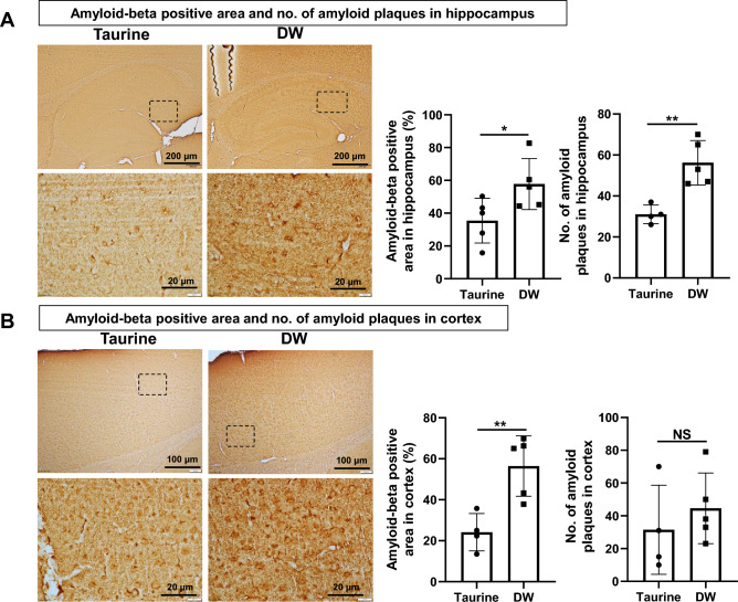
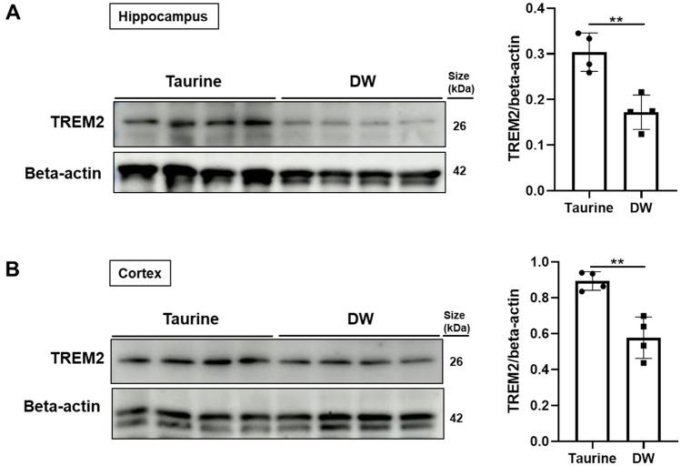
Alzheimer's disease (AD), a chronic neurodegenerative disorder, is the leading cause of dementia. Over-activated microglia is related to amyloid-beta (Aβ) and phosphorylated tau (phospho-tau) accumulation in the AD brain. Taurine is an amino acid with multiple physiological functions including anti-inflammatory effects, and has been reported to be neuroprotective in AD. However, the role of taurine in microglia-mediated AD remains unclear. Here, we examined the effects of taurine on the brains of senescence-accelerated mouse prone 8 (SAMP8) mice by comparing those administered 1% taurine water with those administered distilled water (DW). We observed increased levels of taurine and taurine transporter (TAUT) in the brains of the taurine-treated mice compared with those of control mice. Immunohistochemical and Western blot analyses revealed that taurine significantly reduced the number of activated microglia, levels of phospho-tau and Aβ deposit in the hippocampus and cortex. Triggering receptors expressed on myeloid cells-2 (TREM2) are known to protect against AD pathogenesis. Taurine upregulated TREM2 expression in the hippocampus and cortex. In conclusion, the present study suggests that taurine treatment may upregulate TREM2 to protect against microglia over-activation by decreasing the accumulation of phospho-tau and Aβ; providing an insight into a novel preventive strategy in AD.


Conflict of interest statement

The authors declare no competing interests.

Figures

Figure 1
Figure 1
Effects of taurine treatment on taurine-positive area and TAUT in brain tissue. Representative immunohistochemical staining images of taurine and quantitative analyses of taurine-positive area in the hippocampus (A, top row, bar = 200 µm; bottom row, bar = 50 µm, enlarged from the dashed line box) and cortex (B, top row, bar = 100 µm; bottom row, bar = 50 µm, enlarged from the dashed line box). Taurine-positive areas are visualized in brown. Data are expressed as the means ± SD of 5 hippocampal fields from five SAMP8 mice and 4 specific cortex areas from four SAMP8 mice of each group. Western blots of TAUT expression in the hippocampus (C) and cortex (D). Data are expressed as the means ± SD of quadruplicate samples of each group. *p < 0.05, Student’s t-test.
Figure 2
Figure 2
Effects of taurine treatment on microglial cells in brain tissue. Representative immunohistochemical staining images of Iba-1 (microglia stained in brown) showing microglial activation with quantitative analyses of activated microglia in the hipppocampus (A, top row, bar = 100 µm; bottom row, bar = 20 µm, enlarged from the dashed line box) and cortex (B, top row, bar = 100 µm; bottom row, bar = 20 µm, enlarged from the dashed line box). Nuclei were counterstained with hematoxylin. Quantification of the immunohistochemistry data are expressed as the means ± SD of 3–4 hippocampal fields from three to four SAMP8 mice and 3–4 specific cortex areas from three to four SAMP8 mice of each group. Representative immunohistochemical staining images of TNF-alpha positive area in hippocampus (C, top row, bar = 200 µm; bottom row, bar = 100 µm, enlarged from the dashed line box) and TNF-alpha positive cells in cortex (D, top row, bar = 100 µm; bottom row, bar = 50 µm, enlarged from the dashed line box). Nuclei were counterstained with hematoxylin. Quantification of the immunohistochemistry data are expressed as the means ± SD of 4 hippocampal fields from four SAMP8 mice and 4 specific cortex areas from four SAMP8 mice of each group. *p < 0.05 and **p < 0.01 by Student’s t-test.
Figure 3
Figure 3
Effects of taurine treatment on tau hyperphosphorylation. Representative immunohistochemical staining images of phospho-tau positive area (brown) in the hippocampus (A, top row, bar = 100 µm; bottom row, bar = 20 µm, enlarged from the dashed line box) and cortex (B, top row, bar = 100 µm; bottom row, bar = 20 µm, enlarged from the dashed line box). The rectangular boxes in Fig. 3A indictae the CA1 hippocampal area where phospho-tau was analyzed. Data are expressed as the means ± SD of 3–5 hippocampal fields from three to five SAMP8 mice and 3–5 specific cortex areas from three to five SAMP8 mice of each group. Western blots of PHF1, phospho-tau and total-tau protein levels, bands observed depict the quantitative analyses of PHF1 phospho-tau and total-tau in the hippocampus (C) and cortex (D). Quantification of Western blot data are expressed as the means ± SD of quadruplicate samples of each group. Representative immunohistochemical staining images shows NeuN positive cells in hippocampus (E, top row, bar = 200 µm; bottom row, bar = 100 µm, enlarged from the dashed line box) and cortex (F, top row, bar = 100 µm; bottom row, bar = 50 µm, enlarged from the dashed line box). Data are expressed as the means ± SD of 5 hippocampal fields from five SAMP8 mice and 5 specific cortex areas from five SAMP8 mice of each group. Western blots of NeuN expression in the hippocampus (G) and cortex (H). The values presented are the means ± SD of quadruplicate samples of each group. *p < 0.05, **p < 0.01 and ***p < 0.001 by Student’s t-test.
Figure 4
Figure 4
Effects of taurine treatment on Aβ accumulation in brain tissue. Representative immunohistochemical staining images of Aβ positive area (brown), its quantitative analyses and deposition of amyloid plaques in the hippocampus (A, top row, bar = 200 µm; bottom row, bar = 20 µm, enlarged from the dashed line box) and cortex (B, top row, bar = 100 µm; bottom row, bar = 20 µm, enlarged from the dashed line box). Data are expressed as the means ± SD of 4–5 hippocampal fields from four to five SAMP8 mice and 4–5 specific cortex areas from four to five SAMP8 mice of each group. *p < 0.05 and **p < 0.01, Student’s t-test.
Figure 5
Figure 5
Effects of taurine treatment on TREM2 in brain tissue. Expression of TREM2 were analyzed by Western blotting. Quantitative analyses of TREM2 level in the hippocampus (A) and cortex (B). Quantification of Western blot data is expressed as the means ± SD of quadruplicate samples of each group. **p < 0.01, Student’s t-test.

Similar articles

Cited by

References

    1. Tarawneh R, Holtzman DM. The clinical problem of symptomatic Alzheimer disease and mild cognitive impairment. Cold Spring Harb. Perspect. Med. 2012;2:a006148. doi: 10.1101/cshperspect.a006148. - DOI - PMC - PubMed
    1. Bloom GS. Amyloid-beta and tau: The trigger and bullet in Alzheimer disease pathogenesis. JAMA Neurol. 2014;71:505–508. doi: 10.1001/jamaneurol.2013.5847. - DOI - PubMed
    1. Scheltens P, et al. Alzheimer's disease. Lancet. 2021;397:1577–1590. doi: 10.1016/S0140-6736(20)32205-4. - DOI - PMC - PubMed
    1. Wang Y, Mandelkow E. Tau in physiology and pathology. Nat. Rev. Neurosci. 2016;17:5–21. doi: 10.1038/nrn.2015.1. - DOI - PubMed
    1. Diaz-Aparicio I, et al. Microglia actively remodel adult hippocampal neurogenesis through the phagocytosis secretome. J. Neurosci. 2020;40:1453–1482. doi: 10.1523/JNEUROSCI.0993-19.2019. - DOI - PMC - PubMed