. 2024 Apr;56(4):595-604.
doi: 10.1038/s41588-024-01702-0. Epub 2024 Mar 28.

Cell-type-specific and disease-associated expression quantitative trait loci in the human lung

Affiliations

Cell-type-specific and disease-associated expression quantitative trait loci in the human lung

Heini M Natri et al. Nat Genet. 2024 Apr.

Erratum in

Abstract

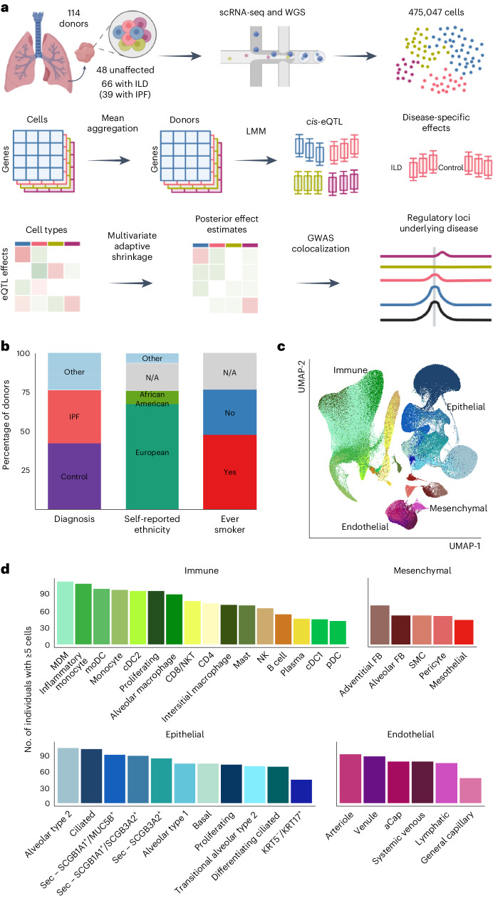
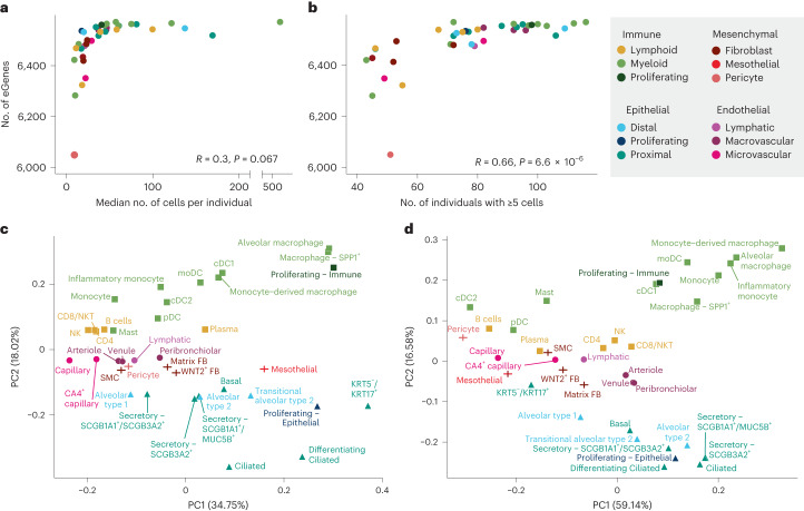
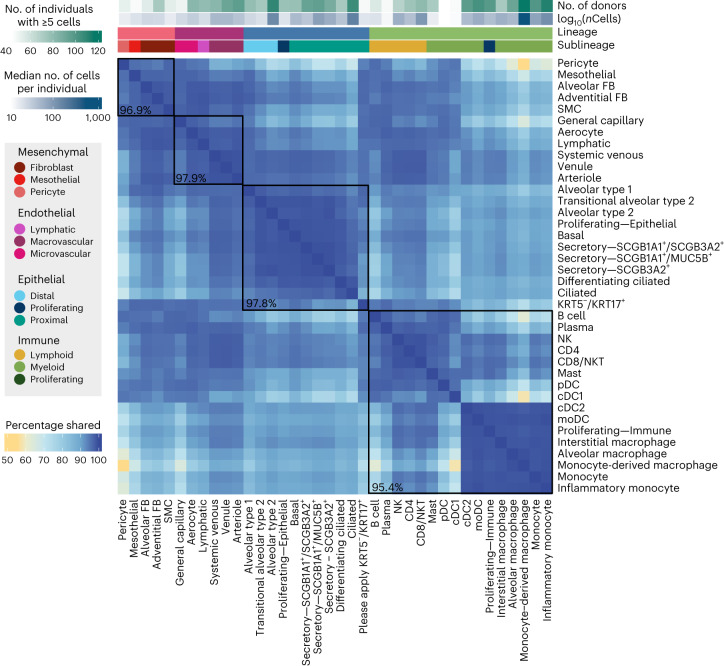
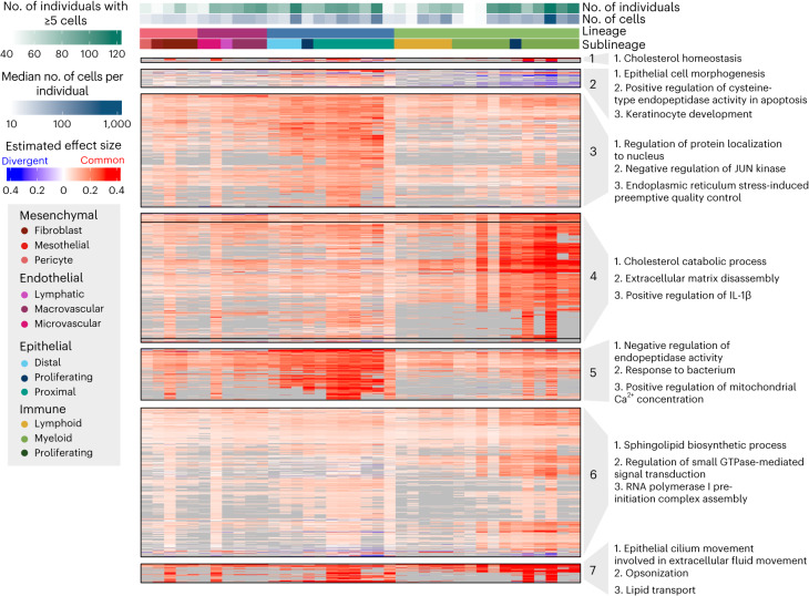
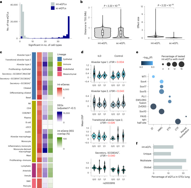
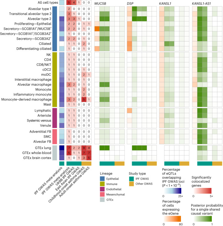
Common genetic variants confer substantial risk for chronic lung diseases, including pulmonary fibrosis. Defining the genetic control of gene expression in a cell-type-specific and context-dependent manner is critical for understanding the mechanisms through which genetic variation influences complex traits and disease pathobiology. To this end, we performed single-cell RNA sequencing of lung tissue from 66 individuals with pulmonary fibrosis and 48 unaffected donors. Using a pseudobulk approach, we mapped expression quantitative trait loci (eQTLs) across 38 cell types, observing both shared and cell-type-specific regulatory effects. Furthermore, we identified disease interaction eQTLs and demonstrated that this class of associations is more likely to be cell-type-specific and linked to cellular dysregulation in pulmonary fibrosis. Finally, we connected lung disease risk variants to their regulatory targets in disease-relevant cell types. These results indicate that cellular context determines the impact of genetic variation on gene expression and implicates context-specific eQTLs as key regulators of lung homeostasis and disease.


Conflict of interest statement

J.A.K. reports advisory board fees from Boehringer Ingelheim, nonfinancial study support from Genentech and grant funding from Boehringer Ingelheim. N.E.B. reports consulting fees from Deepcell. L.B.W. has received advisory board fees from CSL Behring, Quark, Bayer and Merck, and has research contracts with Genentech and CSL Behring. T.S.B. reports consulting fees from Orinove, GRI Bio, Morphic and Novelstar Pharmaceuticals, research grants and contracts from Boehringer Ingelheim and Celgene, and nonfinancial study support from Genentech. R.W. reports consultant fees from Genentech and Boehringer Ingelheim. The remaining authors declare no competing interests.

Figures

Fig. 1
Fig. 1. Mapping eQTLs across cell types in the human lung.
a, Schematic illustration of the present study. b, Percentage proportions of donors according to diagnosis (42.1% unaffected controls, 34.2% IPF, 23.7% other ILD), self-reported ethnicity (66.7% European, 9.6% African American, 17.5% N/A, 6.1% other) and smoking history (46.5% ever smoker, 29.8% never smoker, 23.7% N/A). c, UMAP dimensionality reduction of 437,618 cells across the 38 cell types included in the eQTL analysis. Pseudocoloring indicates cell type; primary cell lineages are labeled. d, Numbers of donors with ≥5 cells for each cell type included in the analysis. LMM, linear mixed model; moDC, monocyte-derived dendritic cell; N/A, not applicable; NK, natural killer cell; NKT, natural killer T cell; pDC, plasmacytoid dendritic cell; SMC, smooth muscle cell. Panel a created with BioRender.com.
Fig. 2
Fig. 2. sc-eQTL structure reflects lineage and cell type relationships.
a, Comparison of the number of eGenes per cell type and the median number of cells per individual of that cell type (two-sided Pearson correlation). Cell types are colored according to sublineage. b, Comparison of the number of eGenes per cell type and the number of individuals with at least five cells of that cell type (Pearson correlation). c, Principal component analysis (PCA) plot of pseudobulk expression across the 6,995 genes included in the eQTL mapping analysis. d, PCA plot of mashr-estimated effect sizes for the top eQTLs (n = 50,389).
Fig. 3
Fig. 3. eQTLs are largely shared between lung cell types.
Percentage of top eQTLs (n = 50,389) shared between two cell types. Top eQTLs are considered shared if they are significant in both cell types (LFSR ≤ 0.1) and the mashr-estimated effect size is within a factor of 0.5. Cell types are annotated above according to lineage, sublineage, the number of individuals with five or more cells and the median number of cells per individual for that cell type. Median pairwise percentage sharing per lineage is shown in black.
Fig. 4
Fig. 4. Multi-cell-type eQTLs act in a highly lineage-specific manner.
Visualization of a representative subset (Methods) of multi-cell-type top eQTLs and IPF-GWAS eQTLs (n = 2,158). eQTLs are clustered according to their estimated effect sizes, with nonsignificant associations set to zero. eQTL effect sizes are not shown (gray) for genes expressed in less than 10% of cells of that cell type. The most common effect direction for each eQTL is shown in red and cell types with opposite effect directions are shown in blue. The top three most significantly enriched GO terms for each cluster, excluding terms with support from less than two genes, are shown.
Fig. 5
Fig. 5. Disease interaction eQTLs converge on pathways relevant to lung fibrosis.
a, Histogram of the cell type sharing of the top int-eQTLs and the top non-int-eQTLs. b, Comparison of absolute distances to the eGene TSS and absolute effect sizes of the top sc-eQTLs (n = 50,506) and int-eQTLs (n = 83,596). Two-sided t-test P values are indicated. In the box plots, the lower and upper hinges correspond to the first and third quartiles. The upper whisker extends from the hinge to the largest value no further than 1.5 times the interquartile range (IQR) from the hinge; the lower whisker extends from the hinge to the smallest value at most 1.5 times the IQR of the hinge. c, Numbers of int-eGenes and differentially expressed genes (DEGs) between fibrotic and unaffected samples, and proportion of their overlap for each cell type included in the int-eQTL analysis. d, Example of an int-eQTL for DSP. In the violin plots, the mean and two s.d. are indicated. e, Top transcription factor motifs enriched among int-eSNPs associated with eGenes that were equally expressed between individuals with ILD and unaffected donors but exhibited differences in eQTL effect sizes. Transcription factors are grouped according to family on the x axis. f, Percentage of int-eQTLs, sc-eQTLs unique to a single cell type, multi-cell-type sc-eQTLs and globally shared sc-eQTLs that are also eQTLs in GTEx lung (P < 1 × 10−6).
Fig. 6
Fig. 6. Cell-type-specific eQTLs colocalize with the lung trait GWAS.
Numbers of SNPs that were nominally significant (P < 1 × 10−6) in the IPF GWAS meta-analysis and also eQTL (blue), the numbers of significant colocalizations between cell type and bulk eQTLs and three IPF GWAS, as well as childhood-onset and adult-onset asthma GWAS (red). Shown are the proportion of cells expressing the gene (orange) and the posterior probabilities for a single shared causal variant between the tested cell types and the GWAS for the selected top IPF-associated genes (MUC5B, DSP, KANSL1, KANSL1-AS1, shown in green) across 27 cell types with at least one colocalized gene.

Update of

References

    1. Umans BD, Battle A, Gilad Y. Where are the disease-associated eQTLs? Trends Genet. 2021;37:109–124. doi: 10.1016/j.tig.2020.08.009. - DOI - PMC - PubMed
    1. Aguet F, et al. The GTEx Consortium atlas of genetic regulatory effects across human tissues. Science. 2020;369:1318–1330. doi: 10.1126/science.aaz1776. - DOI - PMC - PubMed
    1. Lea AJ, Peng J, Ayroles JF. Diverse environmental perturbations reveal the evolution and context-dependency of genetic effects on gene expression levels. Genome Res. 2022;32:1826–1839. - PMC - PubMed
    1. Aguet F, et al. Genetic effects on gene expression across human tissues. Nature. 2017;550:204–213. doi: 10.1038/nature24277. - DOI - PMC - PubMed
    1. Lederer DJ, Martinez FJ. Idiopathic pulmonary fibrosis. N. Engl. J. Med. 2018;378:1811–1823. doi: 10.1056/NEJMra1705751. - DOI - PubMed

MeSH terms