. 2024 Mar 28;24(1):395.
doi: 10.1186/s12885-024-12101-3.

Characterization of tumoricidal activities mediated by a novel immune cell regimen composing interferon-producing killer dendritic cells and tumor-specific cytotoxic T lymphocytes

Affiliations

Characterization of tumoricidal activities mediated by a novel immune cell regimen composing interferon-producing killer dendritic cells and tumor-specific cytotoxic T lymphocytes

Chih-Hao Fang et al. BMC Cancer. .

Abstract

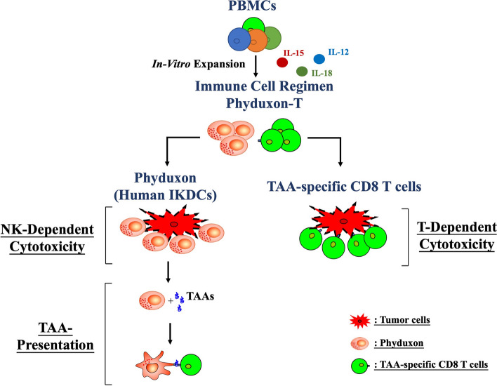
Background: Although immune cell therapy has long been used for treating solid cancer, its efficacy remains limited. Interferon (IFN)-producing killer dendritic cells (IKDCs) exhibit cytotoxicity and present antigens to relevant cells; thus, they can selectively induce tumor-associated antigen (TAA)-specific CD8 T cells and may be useful in cancer treatment. Various protocols have been used to amplify human IKDCs from peripheral sources, but the complexity of the process has prevented their widespread clinical application. Additionally, the induction of TAA-specific CD8 T cells through the adoptive transfer of IKDCs to immunocompromised patients with cancer may be insufficient. Therefore, we developed a method for generating an immune cell-based regimen, Phyduxon-T, comprising a human IKDC counterpart (Phyduxon) and expanded TAA-specific CD8 T cells.

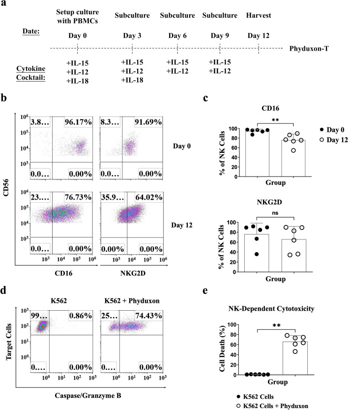
Methods: Peripheral blood mononuclear cells from ovarian cancer patients were cultured with human interleukin (hIL)-15, hIL-12, and hIL-18 to generate Phyduxon-T. Then, its phenotype, cytotoxicity, and antigen-presenting function were evaluated through flow cytometry using specific monoclonal antibodies.

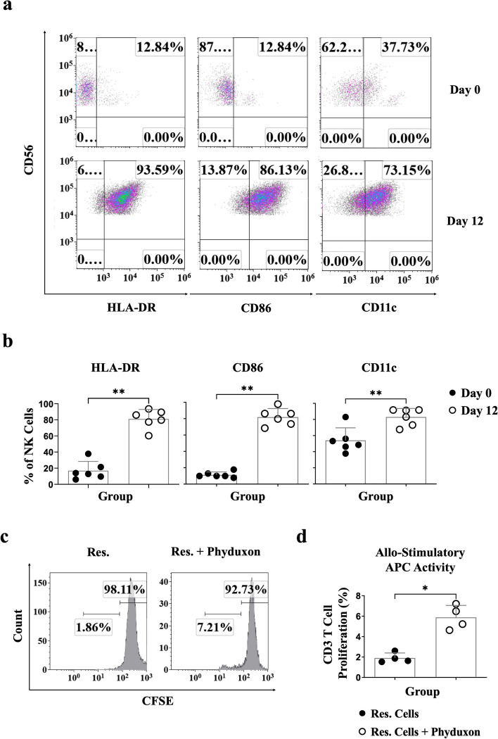
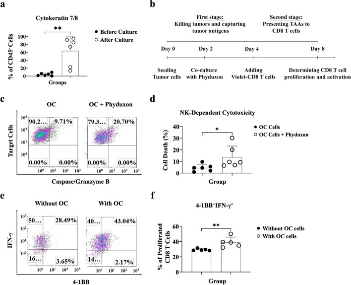
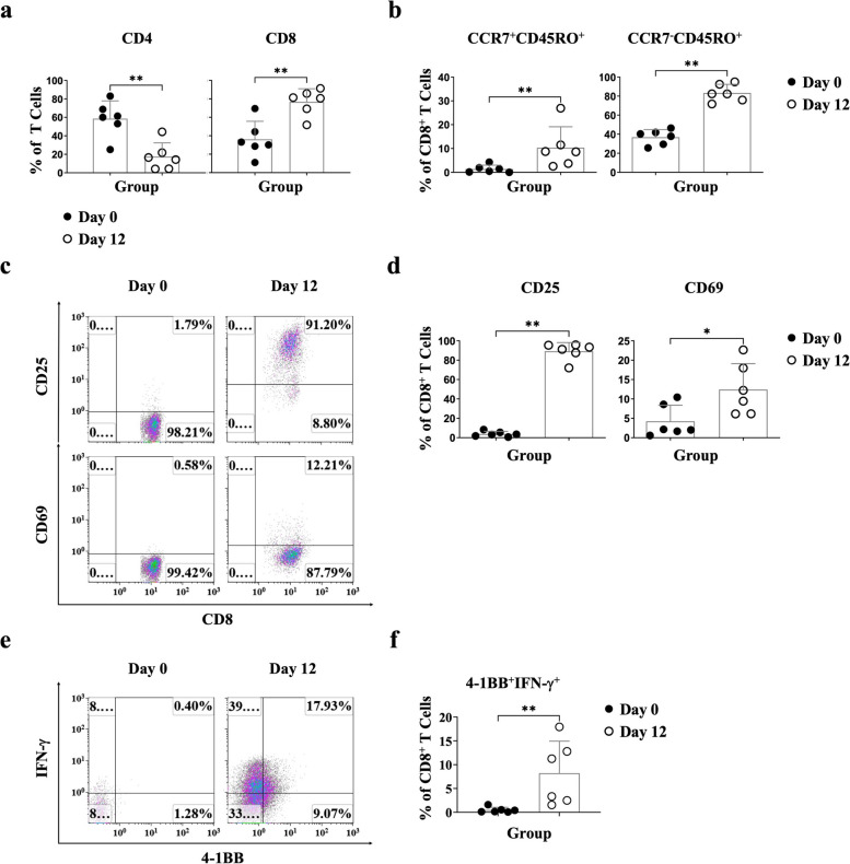
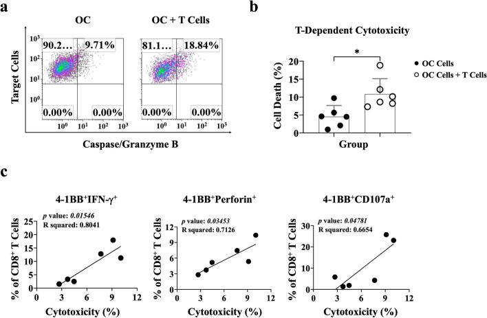
Results: Phyduxon exhibited the characteristics of both natural killer and dendritic cells. This regimen also exhibited cytotoxicity against primary ovarian cancer cells and presented TAAs, thereby inducing TAA-specific CD8 T cells, as evidenced by the expression of 4-1BB and IFN-γ. Notably, the Phyduxon-T manufacturing protocol effectively expanded IFN-γ-producing 4-1BB+ TAA-specific CD8 T cells from peripheral sources; these cells exhibited cytotoxic activities against ovarian cancer cells.

Conclusions: Phyduxon-T, which is a combination of natural killer cells, dendritic cells, and TAA-specific CD8 T cells, may enhance the efficacy of cancer immunotherapy.

Keywords: 4-1BB; IKDCs; Immunotherapy; Phyduxon-T; TAA-specific CD8 T cells.


Conflict of interest statement

J.-M. L., C.-H. F., and Y.-F. C. are employees of FullHope Biomedical Co.,Ltd. They also hold shares in FullHope Biomedical Co.,Ltd. J.-M. L., K.-L. L., C.-H. F., and Y.-F. C. are inventors of patent applications (Application No. 18/170,671). All authors declare no other conflicts of interest related to this study.

Figures

Fig. 1
Fig. 1
Phyduxon, as a human IKDC counterpart, exhibited the phenotypes and activities of NK cells. a Schematic of the manufacturing process for Phyduxon-T. PBMCs were cultured in AIM-V medium plus HPL, containing hIL-15 (days 0, 3, 6, and 9), hIL-12 (days 0, 3, 6, and 9), and hIL-18 (days 0 and 3) for 12 days to generate Phyduxon-T. bc Comparison of the expression of CD16 and NKG2D on CD3CD14CD19CD45+CD56+cells between days 0 and 12 through flow cytometry. d, e Phyduxon was isolated and evaluated for its cytotoxicity after coculturing with K562 tumor cells, analyzing the TFL-4+caspase/granzyme B+ pattern through flow cytometry. The results shown in (b) and (d) are representative of six independent experiments. Data in (c) and (e) are presented as means ± SD of six independent experiments. Differences between groups were analyzed using the nonparametric Mann-Whitney test. p values: **p < 0.01; ns, not significant
Fig. 2
Fig. 2
Phyduxon, as a human IKDC counterpart, exhibited the phenotypes and activities of DCs. a, b Comparison of the expression levels of HLA-DR, CD86, and CD11c on CD3CD14CD19CD45+CD56+cells between days 0 and 12 through flow cytometry. c, d Phyduxon was isolated, and its allostimulatory APC function was evaluated by coculturing with allogeneic CFSE-labeled CD25-responder cells (Res.). Responder CD3 T-cell proliferation was assessed by determining the pattern of CFSE dilution through flow cytometry. The results shown in (a) and (c) are representative of six and four independent experiments, respectively. Data in (b) and (d) are presented as means ± SD of six and four independent experiments, respectively. Differences between groups were analyzed using the nonparametric Mann-Whitney test. values:*p < 0.05,**< 0.01
Fig. 3
Fig. 3
Phyduxon exhibited NK cell-dependent cytotoxicity and induced proliferation and AIM expression of CD8 T cells. a Freshly isolated and cultured OC cells were stained with monoclonal antibodies. The expression of cytokeratin-7/8 on CD45 cells was evaluated through flow cytometry. b A triple-cell coculture assay was performed to determine the APC function of Phyduxon after engaging with primary OC cells. c, d OC cells were cocultured with Phyduxon, and the TFL-4+caspase/granzyme B+ pattern was analyzed through flow cytometry to quantify apoptosis. e, f Phyduxon was cocultured with or without OC cells to determine the expression levels of IFN-γ and 4-1BB on proliferated CD3+CD8+ T cells. The results shown in (c) and (e) are representative of six and five independent experiments, respectively. Data in (a) and (d) are presented as means ± SD of six independent experiments, while data in (f) are presented as means ± SD of five independent experiments. Differences between groups were analyzed using the nonparametric Mann-Whitney test. p values: * p < 0.05, ** p < 0.01
Fig. 4
Fig. 4
Antigen-exposed CD8 T cells were selectively activated and expanded through the Phyduxon-T manufacturing process in vitro. Cells from patients with OC were harvested on day 0 (fresh PBMCs) and day 12 (Phyduxon-T) and stained with monoclonal antibodies to acquire T cell phenotype via flow cytometry. a We evaluated the changes in CD4 and CD8 on T cells, (b) the expression levels of CCR7+CD45RO+(central memory) and CCR7-CD45RO+(effector memory) on CD14CD19CD56TCRγδCD4CD45+TCRαβ+CD8+ cells, (c, d) the expression levels of CD25 and CD69 on CD14CD19CD56TCRγδCD4CD45+CD3+TCRαβ+CD8+cells, (e, f) the expression level of IFN-γ on CD14CD19CD56-CD3+CD8+4-1BB+ cells. The results shown in (c) and (e) are representative of six independent experiments. Data are presented as means ± SD of six independent experiments. Differences between groups were analyzed using the nonparametric Mann-Whitney test. values:*p < 0.05,**p < 0.01
Fig. 5
Fig. 5
AIM+ CD8 T cells exhibited T cell-dependent cytotoxicity against primary epithelial OC. (a, b) OC cells were cocultured with T cells, and the TFL-4+caspase/granzyme B+ pattern was analyzed through flow cytometry to quantify apoptosis. c The correlation between T cell-dependent cytotoxicity and the percentage of 4-1BB+IFN-γ+, 4-1BB+perforin+and 4-1BB+CD107a+on CD8 T cells was determined. The results shown in (a) are representative of six independent experiments. Data are presented as means ± SD of six independent experiments. Differences between groups were analyzed using the nonparametric Mann-Whitney test. values:*p < 0.05
Fig. 6
Fig. 6
Phyduxon-T, as an immune cell-based regimen, exhibits the characteristics of NK cells, DCs, and TAA-specific CD8 T cells. Phyduxon-T is a novel immune cell-based regimen consisting of the human IKDC counterpart, Phyduxon, and TAA-specific CD8 T cells. Phyduxon demonstrates the ability to eliminate tumor cells and present TAAs, thereby inducing TAA-specific CD8 T-cell responses. The manufacturing protocol of Phyduxon-T facilitates the expansion of TAA-specific T cells, enhancing their potential for tumor destruction. In summary, Phyduxon-T exhibits the characteristics of NK cells, DCs, and T cells, mimicking immunosurveillance and providing a new strategy for adoptive cell transfer therapy

Similar articles

Cited by

References

    1. Vakkila J, Lotze MT. Inflammation and necrosis promote tumour growth. Nat Rev Immunol. 2004;4(8):641–648. doi: 10.1038/nri1415. - DOI - PubMed
    1. Bottcher JP, Reis e Sousa C. The role of type 1 conventional dendritic cells in cancer immunity. Trends Cancer. 2018;4(11):784–792. doi: 10.1016/j.trecan.2018.09.001. - DOI - PMC - PubMed
    1. Liu S, Galat V, Galat Y, Lee YKA, Wainwright D, Wu J. NK cell-based cancer immunotherapy: from basic biology to clinical development. J Hematol Oncol. 2021;14(1):7. doi: 10.1186/s13045-020-01014-w. - DOI - PMC - PubMed
    1. Liu J, Fu M, Wang M, Wan D, Wei Y, Wei X. Cancer vaccines as promising immuno-therapeutics: platforms and current progress. J Hematol Oncol. 2022;15(1):28. doi: 10.1186/s13045-022-01247-x. - DOI - PMC - PubMed
    1. Kirtane K, Elmariah H, Chung CH, Abate-Daga D. Adoptive cellular therapy in solid tumor malignancies: review of the literature and challenges ahead. J Immunother Cancer. 2021;9(7):e002723. - PMC - PubMed