

Application of prime editing system to introduce TP53 R248Q hotspot mutation in acute lymphoblastic leukemia cell line
- PMID: 38549229
- PMCID: PMC11145152
- DOI: 10.1111/cas.16162
Application of prime editing system to introduce TP53 R248Q hotspot mutation in acute lymphoblastic leukemia cell line
Abstract
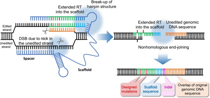
In childhood acute lymphoblastic leukemia (ALL), TP53 gene mutation is associated with chemoresistance in a certain population of relapsed cases. To directly verify the association of TP53 gene mutation with chemoresistance of relapsed childhood ALL cases and improve their prognosis, the development of appropriate human leukemia models having TP53 mutation in the intrinsic gene is required. Here, we sought to introduce R248Q hotspot mutation into the intrinsic TP53 gene in an ALL cell line, 697, by applying a prime editing (PE) system, which is a versatile genome editing technology. The PE2 system uses an artificial fusion of nickase Cas9 and reverse-transcriptase to directly place new genetic information into a target site through a reverse transcriptase template in the prime editing guide RNA (pegRNA). Moreover, in the advanced PE3b system, single guide RNA (sgRNA) matching the edited sequence is also introduced to improve editing efficiency. The initially obtained MDM2 inhibitor-resistant PE3b-transfected subline revealed disrupted p53 transactivation activity, reduced p53 target gene expression, and acquired resistance to chemotherapeutic agents and irradiation. Although the majority of the subline acquired the designed R248Q and adjacent silent mutations, the insertion of the palindromic sequence in the scaffold hairpin structure of pegRNA and the overlap of the original genomic DNA sequence were frequently observed. Targeted next-generation sequencing reconfirmed frequent edit errors in both PE2 and PE3b-transfected 697 cells, and it revealed frequent successful edits in HEK293T cells. These observations suggest a requirement for further modification of the PE2 and PE3b systems for accurate editing in leukemic cells.
Keywords: TP53 gene mutation; acute lymphoblastic leukemia; genome editing; prime editing system; treatment resistance.
© 2024 The Authors. Cancer Science published by John Wiley & Sons Australia, Ltd on behalf of Japanese Cancer Association.
Conflict of interest statement
The authors have no conflict of interest.
Figures


Similar articles
-
Enhancing CRISPR prime editing by reducing misfolded pegRNA interactions.Elife. 2024 Jun 7;12:RP90948. doi: 10.7554/eLife.90948. Elife. 2024. PMID: 38847802 Free PMC article.
-
Prime Editing of Mouse Primary Neurons.Methods Mol Biol. 2025;2910:69-84. doi: 10.1007/978-1-0716-4446-1_5. Methods Mol Biol. 2025. PMID: 40220094
-
Highly efficient generation of isogenic pluripotent stem cell models using prime editing.Elife. 2022 Sep 7;11:e79208. doi: 10.7554/eLife.79208. Elife. 2022. PMID: 36069759 Free PMC article.
-
Prime editing: a search and replace tool with versatile base changes.Yi Chuan. 2022 Nov 20;44(11):993-1008. doi: 10.16288/j.yczz.22-156. Yi Chuan. 2022. PMID: 36384993 Review.
-
Therapeutic Editing of the TP53 Gene: Is CRISPR/Cas9 an Option?Genes (Basel). 2020 Jun 25;11(6):704. doi: 10.3390/genes11060704. Genes (Basel). 2020. PMID: 32630614 Free PMC article. Review.
Cited by
-
CRISPR/Cas9 technology in tumor research and drug development application progress and future prospects.Front Pharmacol. 2025 Jul 8;16:1552741. doi: 10.3389/fphar.2025.1552741. eCollection 2025. Front Pharmacol. 2025. PMID: 40697666 Free PMC article. Review.
-
Overcoming Antigen Escape and T-Cell Exhaustion in CAR-T Therapy for Leukemia.Cells. 2024 Sep 23;13(18):1596. doi: 10.3390/cells13181596. Cells. 2024. PMID: 39329777 Free PMC article. Review.
-
Utility of the Base Editing System for Introducing Drug-Resistant Gene Mutations Into Human Leukemia Cellular Models.Cureus. 2025 Apr 8;17(4):e81889. doi: 10.7759/cureus.81889. eCollection 2025 Apr. Cureus. 2025. PMID: 40342439 Free PMC article.
References
-
- Hof J, Krentz S, van Schewick C, et al. Mutations and deletions of the TP53 gene predict nonresponse to treatment and poor outcome in first relapse of childhood acute lymphoblastic leukemia. J Clin Oncol. 2011;29(23):3185‐3193. - PubMed
-
- Yang F, Brady SW, Tang C, et al. Chemotherapy and mismatch repair deficiency cooperate to fuel TP53 mutagenesis and ALL relapse. Nat Can. 2021;2(8):819‐834. - PubMed
-
- Duffy MJ, Synnott NC, Crown J. Mutant p53 as a target for cancer treatment. Eur J Cancer. 2017;83:258‐265. - PubMed
MeSH terms
Substances
Grants and funding
LinkOut - more resources
Full Text Sources
Research Materials
Miscellaneous
