. 2024 Mar 21:37:191-205.
doi: 10.1016/j.bioactmat.2024.03.008. eCollection 2024 Jul.

Unidirectional gene delivery electrospun fibrous membrane via charge repulsion for tendon repair

Affiliations

Unidirectional gene delivery electrospun fibrous membrane via charge repulsion for tendon repair

Jingwen Liu et al. Bioact Mater. .

Abstract

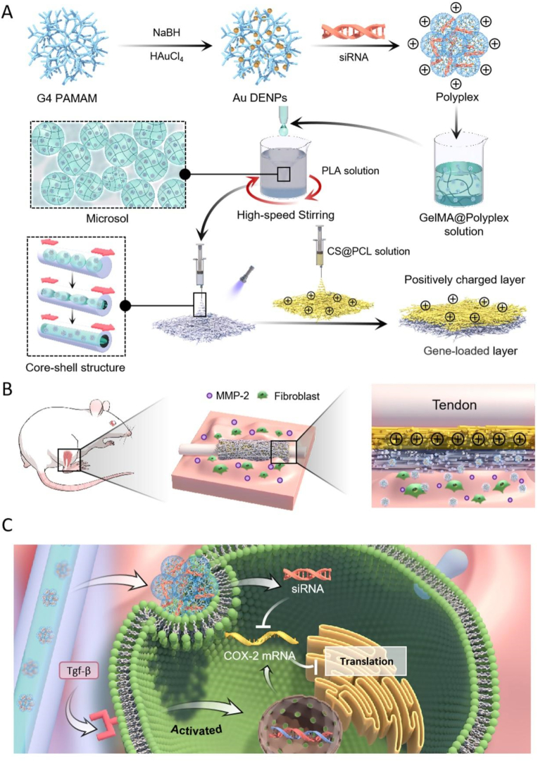
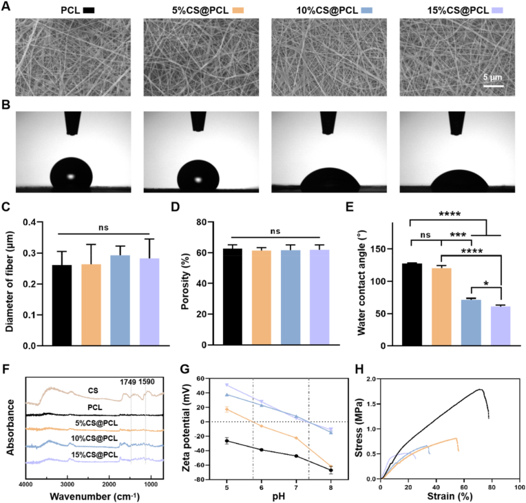
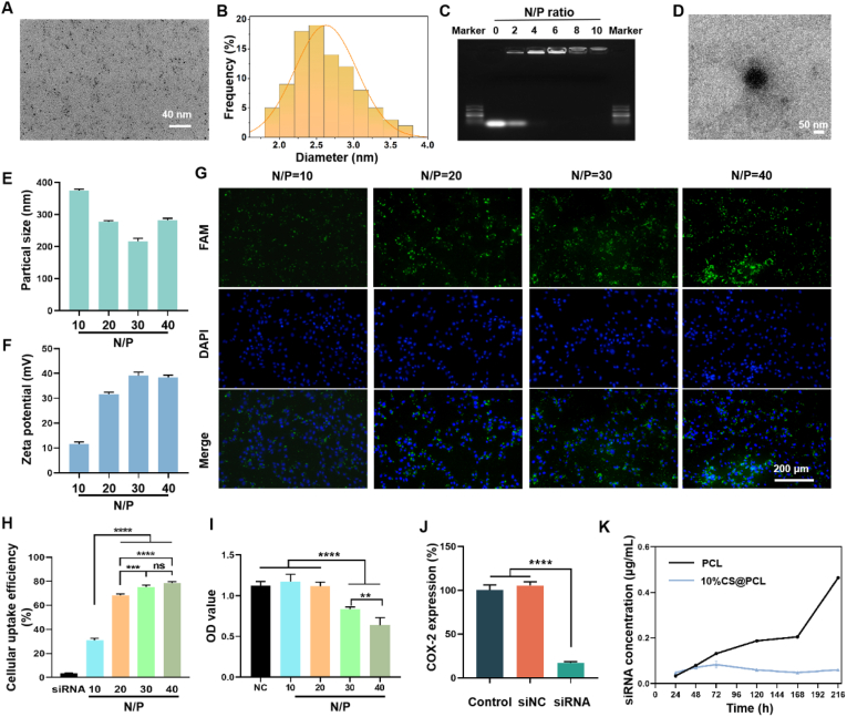
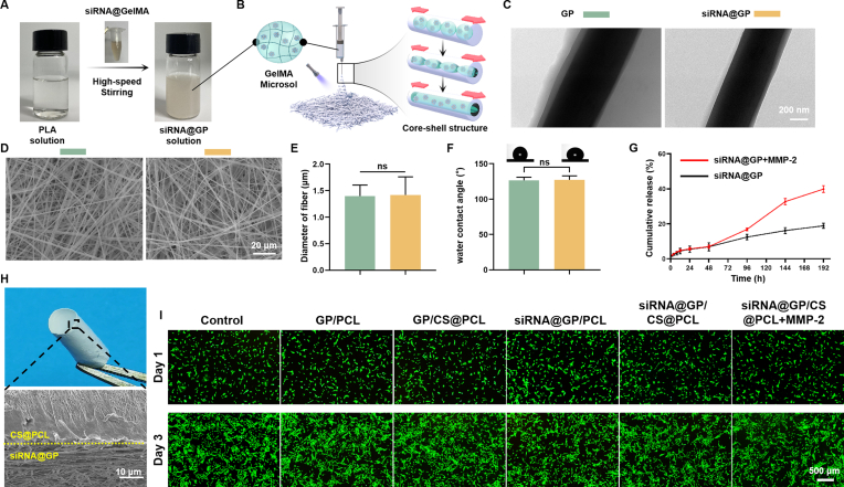
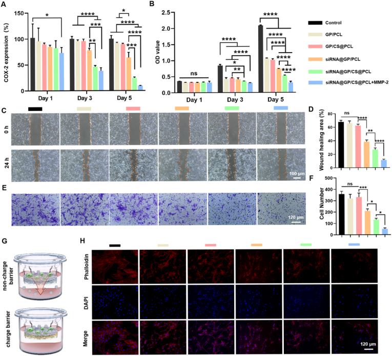
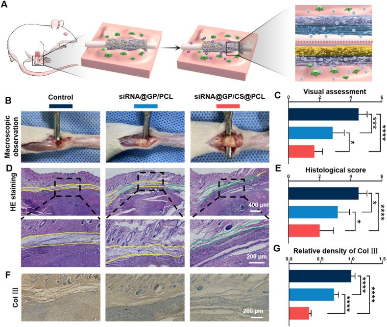
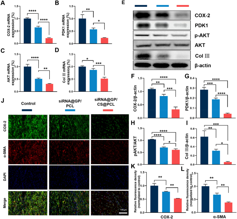
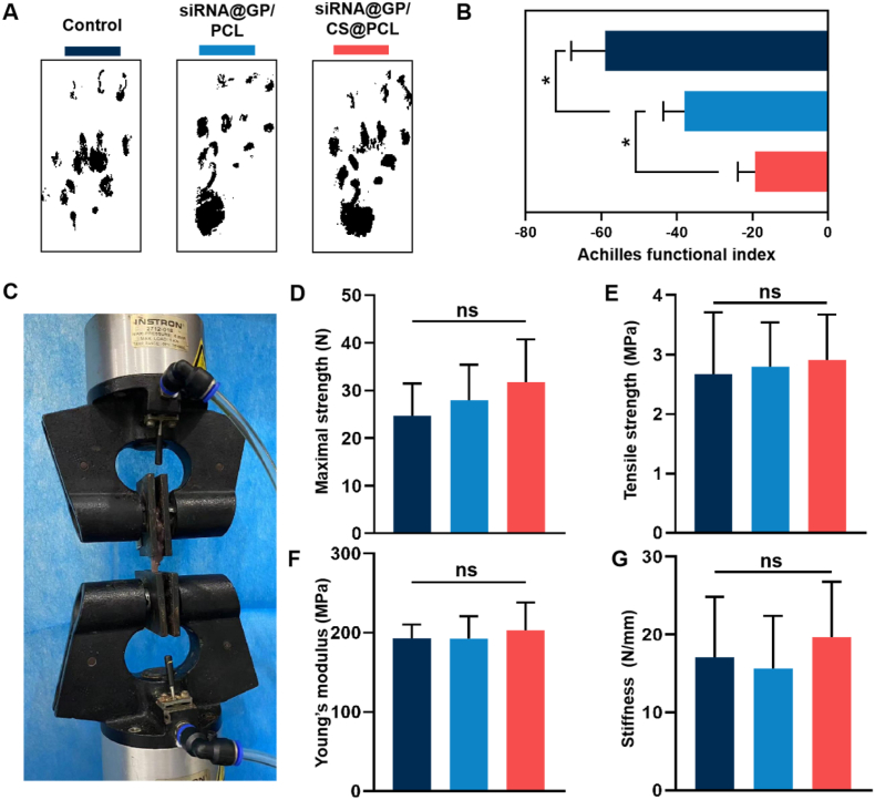
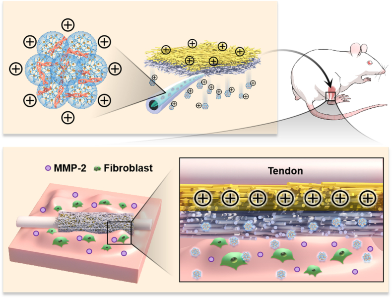
Gene therapy is capable of efficiently regulating the expression of abnormal genes in diseased tissues and expected to be a therapeutic option for refractory diseases. However, unidirectional targeting gene therapy is always desired at the tissue interface. In this study, inspired by the principle that like charges repulse each other, a positively charged micro-nano electrospun fibrous membrane with dual-layer structure was developed by electrospinning technology to achieve unidirectional delivery of siRNA-loaded cationic nanocarriers, thus realizing unidirectional gene therapy at the tendon-paratenon interface. Under the charge repulsion of positively charged layer, more cationic COX-2 siRNA nanocarriers were enriched in peritendinous tissue, which not only improved the bioavailability of the gene drug to prevent the peritendinous adhesion formation, but also avoided adverse effects on the fragile endogenous healing of tendon itself. In summary, this study provides an innovative strategy for unidirectional targeting gene therapy of tissue interface diseases by utilizing charge repulsion to facilitate unidirectional delivery of gene drugs.

Keywords: Charge repulsion; Gene therapy; Tendon repair; Unidirectional delivery.


Conflict of interest statement

The authors declare no conflicts of interest.

Figures

Image 1
Graphical abstract
Fig. 1
Fig. 1
The schematic diagram of the proposed unidirectional gene delivery electrospun fibrous membrane. (A) Schematic representation of the synthesis of siRNA@Au DENPs polyplexes and the fabrication of the dual-layer siRNA@GP/CS@PCL electrospun fibrous membrane consisted of the gene-loaded layer and positively charged layer. (B) Double-layer smart electrospun fibrous membranes were able to respond to the upregulated MMP-2 in the microenvironment, releasing more gene drugs and enriching them in peritendinous tissues via charge repulsion. (C) Inhibits COX-2 synthesis by targeting silencing of COX-2 mRNA, thereby alleviating inflammation.
Fig. 2
Fig. 2
Characterization of the positively charged layer. (A) Representative SEM images of four CS@PCL membranes with different mass ratio. (B) Representative water contact angles images of four CS@PCL membranes with different mass ratio. Quantitative analysis of (C) fiber diameter, (D) porosity analysis and (E) hydrophilicity of four CS@PCL membranes with different mass ratio. (F) FTIR spectra analysis of four CS@PCL membranes with different mass ratio. (G) Surface zeta potential of four CS@PCL with different mass ratios. (H) Tensile properties of four CS@PCL with different mass ratios. (*P < 0.05, ***P < 0.0005, ****P < 0.0001).
Fig. 3
Fig. 3
Characterization of Au DENPs and siRNA@Au DENPs polyplexes. (A) Representative TEM image of Au DENPs. (B) The size distribution of Au DENPs. (C) Agarose gel electrophoresis. (D) Representative TEM image of siRNA@Au DENPs polyplexes. (E) The particle sizes and (F) zeta potentials of siRNA@Au DENPs polyplexes was measured using dynamic light scattering (DLS). (G) Representative fluorescence staining of 208F cells treated with siRNA@Au DENPs with different N/P ratio. (H) Cellular uptake efficiency of siRNA@Au DENPs with different N/P ratio using flow cytometry. (I) Cytotoxicity of siRNA@Au DENPs with different N/P ratio. (J) Gene silencing efficiency of COX-2 siRNA@Au DENPs with N/P ratio = 20. (K) Using Transwell plates to analyze the concentration of siRNA polyplexes of the lower chamber diffused from the upper chamber to verify the charge repulsion effect. (**P < 0.01, ***P < 0.0005, ****P < 0.0001).
Fig. 4
Fig. 4
Construction and Characterization of the gene-loaded layer. (A) After adding siRNA@GelMA solution and stirring at high speed, the clear PLA solution turned into emulsion-like siRNA@GP solution. GP: GelMA@PLA. siRNA@GP: siRNA@GelMA@PLA. (B) The fabrication process of the gene-loaded layer. (C) TEM images of electrospun fibers with core-shell structure. (D) Representative SEM images of the gene-loaded and non-gene-loaded layers. (E) Fiber diameter analysis of the gene-loaded and non-gene-loaded layers. (F) Water contact angle of the gene-loaded and non-gene-loaded layers. (G) siRNA polyplexes released from the gene-loaded layers with or without MMP-2 (0.1 μg/ml). (H) Representative general and cross-sectional SEM image of double-layer membrane. (I) Cytotoxicity detection of different double-layer membranes used in the following in vitro study.
Fig. 5
Fig. 5
Long-lasting gene silencing efficiency, Cell proliferation, Cell migration, and Cell adhesion of unidirectional gene delivery EFMs in vitro. (A) Long-lasting gene silencing efficiency assessment of different double-layer EFMs. (B) Inhibition efficiency of cell proliferation of 208F cells of different double-layer EFMs. (C) Representative images of 208F cells migration after incubation of 0 and 24 h. (D) Quantitative analysis of wound migration rate on the cell migration results. (E) The cell migration was detected by crystal violet staining. (F) Quantitative analysis of 208F cell migration. (G) The schematic representation of anti-adhesion effect of dual-layer EFMs without or with positively charged layer. (H) Fluorescence images of 208F cells seeded on different dual-layer EFMs for 3 days. (*P < 0.05, **P < 0.01, ***P < 0.0005, ****P < 0.0001).
Fig. 6
Fig. 6
Histological analysis of the rat Achilles tendons. (A) Application of dual-layer EFMs without or with positively charged layer for tendon repair. (B) Gross observation of the degree of tendon adhesion on postoperative Day 21. (C) Visual assessment of peritendinous adhesion of the injured tendon. (D) HE staining images (green lines indicate areas without adhesion, yellow lines indicate adhesion areas). (E) Histologic evaluation of peritendinous adhesion. (F) Representative images of immunohistochemical staining of Col Ⅲ. (G) Quantitative analysis of Col Ⅲ expression in peritendinous tissue. (*P < 0.05, ***P < 0.0005, ****P < 0.0001).
Fig. 7
Fig. 7
Assessment of target genes and protein expression in vivo. (A–D) Quantitative qRT-PCR detection of mRNA expression of COX-2, PDK1, AKT and Col Ⅲ. (E) Expression of COX-2, PDK1, p-AKT, AKT and Col Ⅲ in adhesion tissues determined by Western blotting. (F and G) Quantitative analysis of COX-2 and PDK1 levels normalized to β-actin. (H) Quantitative analysis of p-AKT levels normalized to AKT. (I) Quantitative analysis of Col Ⅲ levels normalized to β-actin. (J) Representative Immunofluorescence double-staining images of COX-2 (green) and α-SMA (red) at 3 weeks. (K and L) Semiquantitative analysis on optical density of COX-2 and α-SMA. (*P < 0.05, **P < 0.01, ***P < 0.0005, ****P < 0.0001).
Fig. 8
Fig. 8
Gait tests and Biomechanical testing of the rat Achilles tendon. (A) Gait analysis and (B) Achilles function index (AFI) evaluation of the healed tendon at postoperative Day 21. (C) Biomechanical test of the healed tendon at postoperative Day 21,and Comparison of maximal strength (D), tensile strength (E), Young's modulus (F) and stiffness (G) between the experimental groups.
Fig. 9
Fig. 9
Graphic Abstract. The dual-layer positively charged micro-nano electrospun fibrous membrane with unidirectional gene delivery via charge repulsion and MMP-2 responsiveness was fabricated for tendon repair by unidirectional silencing of COX-2 to modulate the inflammation of peritendinous tissue and on-demand release by responding to up-regulated MMP-2.

Similar articles

Cited by

References

    1. Hurley E.A., Hull D., Shriver S.P. The next phase of human gene-therapy oversight. N. Engl. J. Med. 2019;380:401–402. - PubMed
    1. Alshaer W., Zureigat H., Al Karaki A., Al-Kadash A., Gharaibeh L., Ma'mon M.H., Aljabali A.A., Awidi A. siRNA: mechanism of action, challenges, and therapeutic approaches. Eur. J. Pharmacol. 2021;905:174–178. - PubMed
    1. Paunovska K., Loughrey D., Dahlman J.E. Drug delivery systems for RNA therapeutics. Nat. Rev. Genet. 2022:1–16. - PMC - PubMed
    1. Santos H.S., Rodrigues L., Vera L.N., Poletto E., Filippi-Chiela E., dos Santos Bruschi L.F.R., Schuh R., Baldo G. In situ gene therapy. Curr. Gene Ther. 2021;21:406–430. - PubMed
    1. Tai W., Gao X. Functional peptides for siRNA delivery. Adv. Drug Deliv. Rev. 2017;110:157–168. - PMC - PubMed