Reversible Cerebral Vasoconstriction Syndrome With Typical and Atypical Symptoms: A Case Series
- PMID: 38550478
- PMCID: PMC10977861
- DOI: 10.7759/cureus.55066
Reversible Cerebral Vasoconstriction Syndrome With Typical and Atypical Symptoms: A Case Series
Abstract
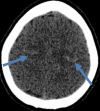
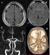
Introduction Reversible cerebral vasoconstriction syndrome (RCVS) is most commonly characterized by focal or diffuse severe thunderclap headache with or without focal neurological deficits and associated transient focal vasoconstriction of the intracerebral arteries lasting up to three months. We present six patients diagnosed as RCVS, with three patients presenting with focal neurological deficits without headache and the remaining three with severe headache alone. Neuroimaging revealed focal subarachnoid bleed with or without segmental intracerebral vasospasm, which resolved over three months. Despite thunderclap headache being the most prevalent symptom associated with RCVS, the absence of this symptom should not preclude the diagnosis, especially in the presence of cortical subarachnoid hemorrhage (SAH) or focal segmental intracerebral arterial narrowing. Methods This case series is a retrospective analysis of all patients diagnosed with RCVS between 2018 and 2022, focusing on clinical symptoms, imaging findings, and management. Results Six patients (three males and three females) were diagnosed with RCVS between 2018 and 2022. Three patients presented with typical symptoms, while the remaining three presented with atypical symptoms. Neuroimaging findings ranged from normal to focal SAH with or without arterial narrowing. Conclusion This case series underscores the diverse clinical presentations of RCVS, emphasizing that while thunderclap headache is the predominant symptom, its absence should not exclude the possibility of RCVS, especially when accompanied by focal neurological deficits or cortical SAH. Neuroimaging played a crucial role in identifying the spectrum of findings. These findings highlight the importance of comprehensive evaluation and consideration of RCVS in patients presenting with neurological symptoms, even in the absence of typical headache features.
Keywords: ct; ct angio; mri; primary angiitis of the central nervous system; reversible cerebral vasoconstriction syndrome; subarachnoid haemorrhage; thunderclap headache.
Copyright © 2024, Sivagurunathan et al.
Conflict of interest statement
The authors have declared that no competing interests exist.
Figures


Similar articles
-
Reversible cerebral vasoconstriction syndromes presenting with subarachnoid hemorrhage: a case series.J Neurointerv Surg. 2011 Sep;3(3):272-8. doi: 10.1136/jnis.2010.004242. Epub 2011 Feb 4. J Neurointerv Surg. 2011. PMID: 21990840
-
Reversible cerebral vasoconstriction syndrome or primary angiitis of the central nervous system?Can J Neurol Sci. 2007 Nov;34(4):467-77. Can J Neurol Sci. 2007. PMID: 18062458
-
Reversible cerebral vasoconstriction syndrome as a cause of thunderclap headache: a retrospective case series study.Am J Emerg Med. 2015 Jun;33(6):859.e3-6. doi: 10.1016/j.ajem.2014.12.026. Epub 2014 Dec 19. Am J Emerg Med. 2015. PMID: 25583268
-
Reversible Cerebral Vasoconstriction Syndrome Without Typical Thunderclap Headache.Headache. 2016 Apr;56(4):674-87. doi: 10.1111/head.12794. Epub 2016 Mar 26. Headache. 2016. PMID: 27016378 Review.
-
Reversible cerebral vasoconstriction syndrome: review of neuroimaging findings.Radiol Med. 2022 Sep;127(9):981-990. doi: 10.1007/s11547-022-01532-2. Epub 2022 Aug 6. Radiol Med. 2022. PMID: 35932443 Free PMC article. Review.
References
-
- Narrative review: reversible cerebral vasoconstriction syndromes. Calabrese LH, Dodick DW, Schwedt TJ, Singhal AB. Ann Intern Med. 2007;146:34–44. - PubMed
-
- Postpartum angiopathy and other cerebral vasoconstriction syndromes. Singhal AB, Bernstein RA. Neurocrit Care. 2005;3:91–97. - PubMed
-
- Thunderclap headache. Ducros A, Bousser MG. BMJ. 2013;346:0. - PubMed
-
- The clinical and radiological spectrum of reversible cerebral vasoconstriction syndrome. A prospective series of 67 patients. Ducros A, Boukobza M, Porcher R, Sarov M, Valade D, Bousser MG. Brain. 2007;130:3091–3101. - PubMed
LinkOut - more resources
Full Text Sources
