Review
. 2024 Mar 14:15:1374796.
doi: 10.3389/fimmu.2024.1374796. eCollection 2024.

Mitochondrial extracellular vesicles, autoimmunity and myocarditis

Affiliations
Review

Mitochondrial extracellular vesicles, autoimmunity and myocarditis

Damian N Di Florio et al. Front Immunol. .

Abstract

For many decades viral infections have been suspected as 'triggers' of autoimmune disease, but mechanisms for how this could occur have been difficult to establish. Recent studies have shown that viral infections that are commonly associated with viral myocarditis and other autoimmune diseases such as coxsackievirus B3 (CVB3) and SARS-CoV-2 target mitochondria and are released from cells in mitochondrial vesicles that are able to activate the innate immune response. Studies have shown that Toll-like receptor (TLR)4 and the inflammasome pathway are activated by mitochondrial components. Autoreactivity against cardiac myosin and heart-specific immune responses that occur after infection with viruses where the heart is not the primary site of infection (e.g., CVB3, SARS-CoV-2) may occur because the heart has the highest density of mitochondria in the body. Evidence exists for autoantibodies against mitochondrial antigens in patients with myocarditis and dilated cardiomyopathy. Defects in tolerance mechanisms like autoimmune regulator gene (AIRE) may further increase the likelihood of autoreactivity against mitochondrial antigens leading to autoimmune disease. The focus of this review is to summarize current literature regarding the role of viral infection in the production of extracellular vesicles containing mitochondria and virus and the development of myocarditis.

Keywords: AIRE; autoimmune disease; coxsackievirus; extracellular vesicles; mitochondria; mitochondrial-derived vesicles; myocarditis.


Conflict of interest statement

The authors declare that the research was conducted in the absence of any commercial or financial relationships that could be construed as a potential conflict of interest. The author(s) declared that they were an editorial board member of Frontiers, at the time of submission. This had no impact on the peer review process and the final decision.

Figures

Figure 1
Figure 1
Mitochondrial derived vesicles (MDVs) from cardiac mitochondria. Widefield transmission electron microscopic images of mitochondria isolated from bovine heart. (A) 60-100 nm vesicles containing mitochondria. Scale bar, 500 nm, (B) MDV budding from mitochondria containing inner and outer mitochondrial membrane, (C) Protrusion of MDV from mitochondria showing constriction at its base, (D) MDV forming with only outer mitochondrial membrane. Panels (B–D), scale bars 100 nm. Reused with permission from (54).
Figure 2
Figure 2
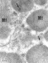
CVB3 localizes around and within murine cardiac mitochondria during myocarditis. Immunogold electron micrograph of mouse cardiomyocyte with CVB3 myocarditis on day 8 post infection. Black dots (arrows) are gold staining of CVB3 viral genome localizing around and in cardiomyocyte mitochondria. Scale bar, 100 nm. Mt, mitochondria; Mf, myofibril. Reused with permission from (58).
Figure 3
Figure 3
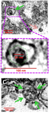
CVB3 identified in EVs using transmission electron microscopy. (A) Widefield transmission electron microscopic view of single virion (green arrow) in an extracellular EV or free virion (pink arrow) from culture of CVB3 in C2C12 cells. (B) Higher digital magnification (dashed purple box) of a virus-like particle revealed an icosahedral shape structure (dashed green polygon) slightly larger than 31 nm in diameter enclosed within a membrane structure. (C) Large EV containing multiple virions (green arrows). Scale bars = 100 nm. Reused with permission from (47).
Figure 4
Figure 4
Formation of mitochondrial EVs from cardiomyocytes after CVB3 infection (1). CVB3 gains entry to cardiomyocytes via the coxsackievirus adrenoreceptor (CAR) or passive entry from previously formed mitochondrial EVs containing replicative virus (2). CVB3 mitochondrial localization induces mitochondrial stress and damage leading to (3a) mitochondrial-derived vesicle (MDV) formation and Drp1-mediated mitochondrial fission and recruitment of the endoplasmic reticulum (ER) for autophagosome formation alongside LC3 lipidation (LC3-II). (3b) MDVs containing replicative and or non-replicative viral particles may either eject from the cell or join multi-vesicular bodies before release from cardiomyocytes (MDVs can also be slated for receptor mediated mitophagy and potentially escape the cell without GABARAPL phosphorylation, which is not shown in this diagram) (4). LC3-II binds mitophagy adaptors situated on the outer mitochondrial membrane to form a mitophagosome with GABARAPL proteins on the endoplasmic reticulum (ER) facing the cytosol (5). Phosphorylation of the mitophagosome on GABARAPL proteins by tank-binding kinase 1 (TBK1) leads mitophagosomes to subsequent (6) lysosomal fusion and degradation (7). Non-phosphorylated mitophagosomes do not proceed to fusion with the lysosome but either (8) join the multivesicular body for cell release and dissemination or are ejected alone. The resulting EVs containing mitochondrial components and viral particles (replicative and/or non-replicative) we term as “mitopods” or mitochondrial escape-pods for CVB3. The two major sources of mitopods are MDV-derived or fission-derived. Another possible distinguishing feature of fission-derived versus MDV-derived mitopods would be an additional membrane derived from the ER. This figure was created using BioRender.

References

    1. Sogkas G, Atschekzei F, Adriawan IR, Dubrowinskaja N, Witte T, Schmidt RE. Cellular and molecular mechanisms breaking immune tolerance in inborn errors of immunity. Cell Mol Immunol. (2021) 18:1122–40. doi: 10.1038/s41423-020-00626-z - DOI - PMC - PubMed
    1. Fairweather D, Kaya Z, Shellam GR, Lawson CM, Rose NR. From infection to autoimmunity. J Autoimmun. (2001) 16:175–86. doi: 10.1006/jaut.2000.0492 - DOI - PubMed
    1. Shamim EA, Miller FW. Familial autoimmunity and the idiopathic inflammatory myopathies. Curr Rheumatol Rep. (2000) 2:201–11. doi: 10.1007/s11926-000-0080-0 - DOI - PubMed
    1. Fairweather D, Root-Bernstein R. Autoimmune disease: Mechanism. In: Encyclopedia of life sciences. John Wiley & Sons, Ltd, Chichester, UK: (2015). p. 1–7. doi: 10.1002/9780470015902.a0020193.pub2 - DOI
    1. Fairweather D, Rose NR. Type 1 diabetes: Virus infection or autoimmune disease? Nat Immunol. (2002) 3:338–40. doi: 10.1038/ni0402-338 - DOI - PubMed

Publication types